Zinc in PDB 7mc9: X-Ray Structure of Pedv Papain-Like Protease 2 Bound to Ub-Pa
Enzymatic activity of X-Ray Structure of Pedv Papain-Like Protease 2 Bound to Ub-Pa
All present enzymatic activity of X-Ray Structure of Pedv Papain-Like Protease 2 Bound to Ub-Pa:
3.4.19.12;
3.6.4.12;
3.6.4.13;
Protein crystallography data
The structure of X-Ray Structure of Pedv Papain-Like Protease 2 Bound to Ub-Pa, PDB code: 7mc9
was solved by
I.A.Durie,
J.V.Dzimianski,
C.M.Daczkowski,
S.D.Pegan,
with X-Ray Crystallography technique. A brief refinement statistics is given in the table below:
Resolution Low / High (Å)
|
49.07 /
3.10
|
Space group
|
P 21 21 21
|
Cell size a, b, c (Å), α, β, γ (°)
|
98.138,
136.873,
193.348,
90,
90,
90
|
R / Rfree (%)
|
20 /
25.7
|
Zinc Binding Sites:
Pages:
>>> Page 1 <<<
Page 2, Binding sites: 11 -
14;
Binding sites:
The binding sites of Zinc atom in the X-Ray Structure of Pedv Papain-Like Protease 2 Bound to Ub-Pa
(pdb code 7mc9). This binding sites where shown within
5.0 Angstroms radius around Zinc atom.
In total 14 binding sites of Zinc where determined in the
X-Ray Structure of Pedv Papain-Like Protease 2 Bound to Ub-Pa, PDB code: 7mc9:
Jump to Zinc binding site number:
1;
2;
3;
4;
5;
6;
7;
8;
9;
10;
Zinc binding site 1 out
of 14 in 7mc9
Go back to
Zinc Binding Sites List in 7mc9
Zinc binding site 1 out
of 14 in the X-Ray Structure of Pedv Papain-Like Protease 2 Bound to Ub-Pa
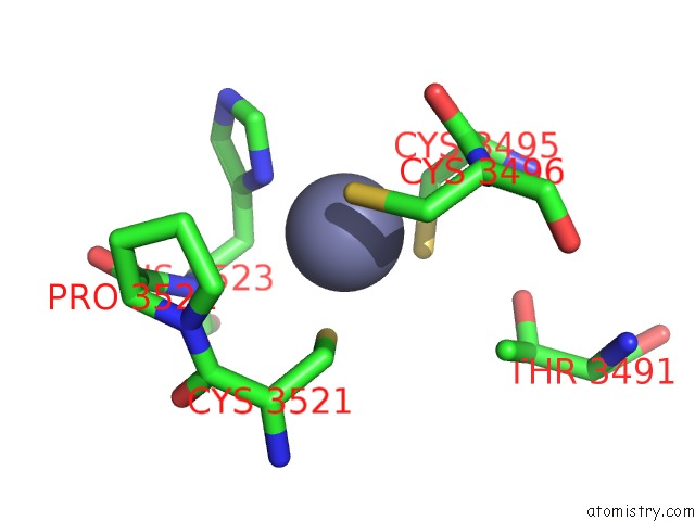
Mono view
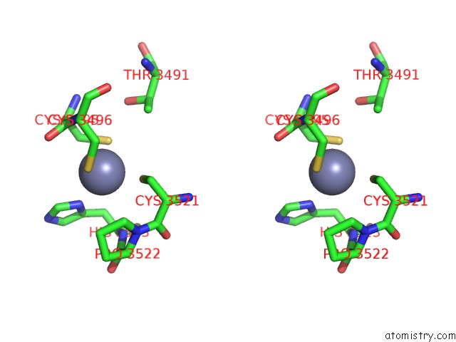
Stereo pair view
|
A full contact list of Zinc with other atoms in the Zn binding
site number 1 of X-Ray Structure of Pedv Papain-Like Protease 2 Bound to Ub-Pa within 5.0Å range:
probe
|
atom
|
residue
|
distance (Å)
|
B
|
Occ
|
A:Zn3701
b:52.7
occ:1.00
|
SG
|
A:CYS3495
|
2.3
|
81.2
|
1.0
|
ND1
|
A:HIS3523
|
2.4
|
66.1
|
1.0
|
SG
|
A:CYS3496
|
2.4
|
46.1
|
1.0
|
SG
|
A:CYS3521
|
2.5
|
45.5
|
1.0
|
CB
|
A:CYS3495
|
3.1
|
50.7
|
1.0
|
CG
|
A:HIS3523
|
3.2
|
53.3
|
1.0
|
CB
|
A:CYS3521
|
3.2
|
49.7
|
1.0
|
CE1
|
A:HIS3523
|
3.4
|
68.6
|
1.0
|
CB
|
A:HIS3523
|
3.4
|
48.4
|
1.0
|
C
|
A:CYS3495
|
3.6
|
70.4
|
1.0
|
CB
|
A:CYS3496
|
3.6
|
57.0
|
1.0
|
N
|
A:CYS3496
|
3.7
|
59.9
|
1.0
|
O
|
A:CYS3495
|
3.9
|
70.7
|
1.0
|
CA
|
A:CYS3495
|
3.9
|
65.2
|
1.0
|
N
|
A:HIS3523
|
4.0
|
49.6
|
1.0
|
CA
|
A:CYS3496
|
4.3
|
50.8
|
1.0
|
CA
|
A:HIS3523
|
4.3
|
48.5
|
1.0
|
CD2
|
A:HIS3523
|
4.3
|
53.9
|
1.0
|
NE2
|
A:HIS3523
|
4.4
|
61.5
|
1.0
|
CD
|
A:PRO3522
|
4.4
|
57.2
|
1.0
|
N
|
A:PRO3522
|
4.5
|
55.5
|
1.0
|
CA
|
A:CYS3521
|
4.5
|
45.5
|
1.0
|
CG
|
A:PRO3522
|
4.6
|
58.9
|
1.0
|
C
|
A:CYS3521
|
4.6
|
52.2
|
1.0
|
CG2
|
A:THR3491
|
4.6
|
51.7
|
1.0
|
OG1
|
A:THR3491
|
4.8
|
60.4
|
1.0
|
N
|
A:CYS3495
|
4.9
|
60.4
|
1.0
|
C
|
A:PRO3522
|
4.9
|
49.1
|
1.0
|
|
Zinc binding site 2 out
of 14 in 7mc9
Go back to
Zinc Binding Sites List in 7mc9
Zinc binding site 2 out
of 14 in the X-Ray Structure of Pedv Papain-Like Protease 2 Bound to Ub-Pa
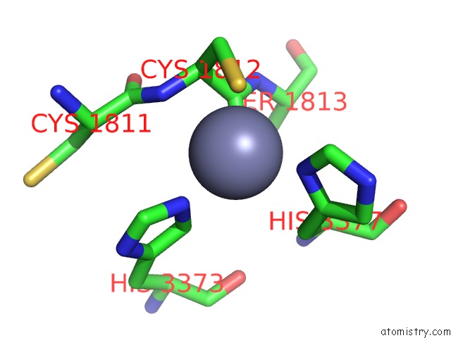
Mono view
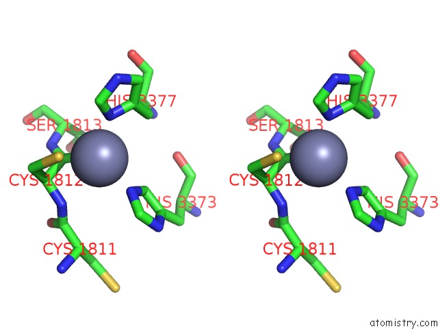
Stereo pair view
|
A full contact list of Zinc with other atoms in the Zn binding
site number 2 of X-Ray Structure of Pedv Papain-Like Protease 2 Bound to Ub-Pa within 5.0Å range:
probe
|
atom
|
residue
|
distance (Å)
|
B
|
Occ
|
A:Zn3702
b:53.0
occ:1.00
|
O
|
O:CYS1812
|
2.0
|
48.0
|
1.0
|
ND1
|
A:HIS3377
|
2.1
|
35.5
|
1.0
|
SG
|
O:CYS1812
|
2.2
|
40.4
|
1.0
|
NE2
|
A:HIS3373
|
2.3
|
58.5
|
1.0
|
C
|
O:CYS1812
|
2.8
|
47.0
|
1.0
|
CE1
|
A:HIS3377
|
2.9
|
38.0
|
1.0
|
N
|
O:CYS1812
|
3.1
|
62.6
|
1.0
|
CG
|
A:HIS3377
|
3.2
|
36.5
|
1.0
|
CD2
|
A:HIS3373
|
3.2
|
58.4
|
1.0
|
CA
|
O:CYS1812
|
3.2
|
52.7
|
1.0
|
CB
|
O:CYS1812
|
3.2
|
44.1
|
1.0
|
CE1
|
A:HIS3373
|
3.3
|
67.0
|
1.0
|
CB
|
A:HIS3377
|
3.6
|
43.5
|
1.0
|
NE2
|
A:HIS3377
|
4.0
|
37.2
|
1.0
|
N
|
O:SER1813
|
4.0
|
53.0
|
1.0
|
CD2
|
A:HIS3377
|
4.2
|
34.9
|
1.0
|
C
|
O:CYS1811
|
4.2
|
60.5
|
1.0
|
CG
|
A:HIS3373
|
4.4
|
69.9
|
1.0
|
ND1
|
A:HIS3373
|
4.4
|
75.0
|
1.0
|
CA
|
O:SER1813
|
4.6
|
46.6
|
1.0
|
CA
|
A:HIS3377
|
4.6
|
45.6
|
1.0
|
CA
|
O:CYS1811
|
4.8
|
63.7
|
1.0
|
|
Zinc binding site 3 out
of 14 in 7mc9
Go back to
Zinc Binding Sites List in 7mc9
Zinc binding site 3 out
of 14 in the X-Ray Structure of Pedv Papain-Like Protease 2 Bound to Ub-Pa
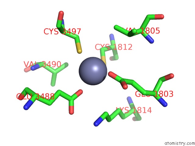
Mono view
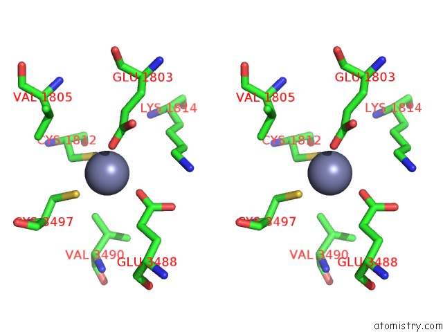
Stereo pair view
|
A full contact list of Zinc with other atoms in the Zn binding
site number 3 of X-Ray Structure of Pedv Papain-Like Protease 2 Bound to Ub-Pa within 5.0Å range:
probe
|
atom
|
residue
|
distance (Å)
|
B
|
Occ
|
A:Zn3703
b:140.3
occ:1.00
|
SG
|
C:CYS1812
|
2.3
|
83.3
|
1.0
|
SG
|
A:CYS3497
|
2.4
|
93.3
|
1.0
|
OE2
|
A:GLU3488
|
2.4
|
91.6
|
1.0
|
OE2
|
C:GLU1803
|
2.4
|
88.3
|
1.0
|
CD
|
A:GLU3488
|
3.5
|
71.2
|
1.0
|
CD
|
C:GLU1803
|
3.5
|
75.8
|
1.0
|
CB
|
C:CYS1812
|
3.7
|
63.4
|
1.0
|
CB
|
A:CYS3497
|
3.8
|
67.9
|
1.0
|
CG
|
A:GLU3488
|
3.9
|
61.5
|
1.0
|
CG
|
C:GLU1803
|
4.0
|
61.5
|
1.0
|
CE
|
C:LYS1814
|
4.4
|
67.7
|
1.0
|
OE1
|
A:GLU3488
|
4.5
|
77.1
|
1.0
|
OE1
|
C:GLU1803
|
4.5
|
78.9
|
1.0
|
CG2
|
A:VAL3490
|
4.8
|
63.7
|
1.0
|
CG2
|
C:VAL1805
|
4.8
|
53.3
|
1.0
|
|
Zinc binding site 4 out
of 14 in 7mc9
Go back to
Zinc Binding Sites List in 7mc9
Zinc binding site 4 out
of 14 in the X-Ray Structure of Pedv Papain-Like Protease 2 Bound to Ub-Pa
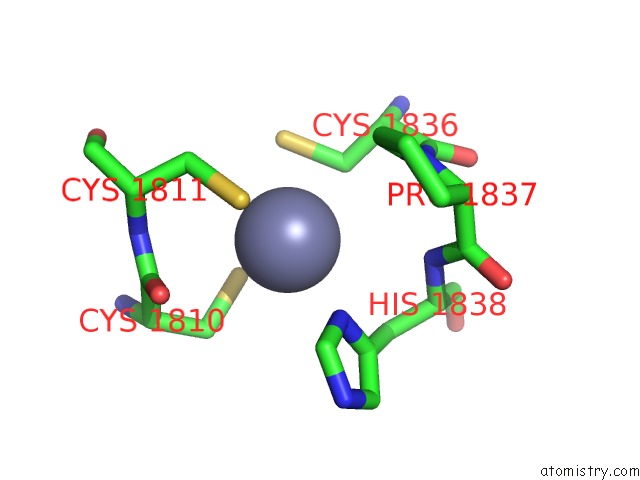
Mono view
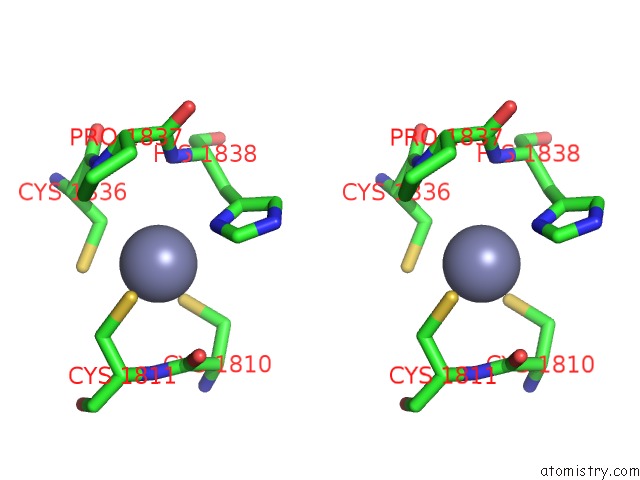
Stereo pair view
|
A full contact list of Zinc with other atoms in the Zn binding
site number 4 of X-Ray Structure of Pedv Papain-Like Protease 2 Bound to Ub-Pa within 5.0Å range:
probe
|
atom
|
residue
|
distance (Å)
|
B
|
Occ
|
C:Zn2001
b:36.2
occ:1.00
|
SG
|
C:CYS1810
|
2.4
|
60.6
|
1.0
|
SG
|
C:CYS1811
|
2.6
|
55.1
|
1.0
|
ND1
|
C:HIS1838
|
2.8
|
55.4
|
1.0
|
CB
|
C:CYS1836
|
2.8
|
52.8
|
1.0
|
SG
|
C:CYS1836
|
3.1
|
119.6
|
1.0
|
CB
|
C:CYS1810
|
3.4
|
60.8
|
1.0
|
CB
|
C:CYS1811
|
3.5
|
64.8
|
1.0
|
CG
|
C:HIS1838
|
3.6
|
40.3
|
1.0
|
CB
|
C:HIS1838
|
3.7
|
48.7
|
1.0
|
C
|
C:CYS1810
|
3.7
|
75.5
|
1.0
|
CE1
|
C:HIS1838
|
3.7
|
58.6
|
1.0
|
N
|
C:HIS1838
|
3.8
|
55.9
|
1.0
|
N
|
C:CYS1811
|
3.9
|
66.2
|
1.0
|
O
|
C:CYS1810
|
3.9
|
79.2
|
1.0
|
CD
|
C:PRO1837
|
4.1
|
61.5
|
1.0
|
CA
|
C:CYS1836
|
4.1
|
61.3
|
1.0
|
N
|
C:PRO1837
|
4.2
|
57.4
|
1.0
|
CA
|
C:CYS1810
|
4.2
|
61.8
|
1.0
|
CA
|
C:CYS1811
|
4.3
|
57.8
|
1.0
|
CA
|
C:HIS1838
|
4.4
|
53.7
|
1.0
|
C
|
C:CYS1836
|
4.4
|
60.9
|
1.0
|
CD2
|
C:HIS1838
|
4.7
|
50.0
|
1.0
|
C
|
C:PRO1837
|
4.7
|
58.2
|
1.0
|
NE2
|
C:HIS1838
|
4.8
|
57.7
|
1.0
|
CA
|
C:PRO1837
|
4.9
|
55.1
|
1.0
|
|
Zinc binding site 5 out
of 14 in 7mc9
Go back to
Zinc Binding Sites List in 7mc9
Zinc binding site 5 out
of 14 in the X-Ray Structure of Pedv Papain-Like Protease 2 Bound to Ub-Pa
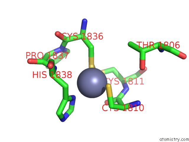
Mono view
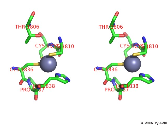
Stereo pair view
|
A full contact list of Zinc with other atoms in the Zn binding
site number 5 of X-Ray Structure of Pedv Papain-Like Protease 2 Bound to Ub-Pa within 5.0Å range:
probe
|
atom
|
residue
|
distance (Å)
|
B
|
Occ
|
E:Zn2001
b:68.3
occ:1.00
|
SG
|
E:CYS1836
|
1.8
|
49.4
|
1.0
|
ND1
|
E:HIS1838
|
2.2
|
67.7
|
1.0
|
SG
|
E:CYS1810
|
2.2
|
61.0
|
1.0
|
SG
|
E:CYS1811
|
2.6
|
66.5
|
1.0
|
O
|
E:CYS1810
|
2.9
|
77.8
|
1.0
|
CB
|
E:CYS1810
|
2.9
|
61.7
|
1.0
|
CG
|
E:HIS1838
|
3.0
|
54.3
|
1.0
|
C
|
E:CYS1810
|
3.1
|
70.1
|
1.0
|
CE1
|
E:HIS1838
|
3.2
|
63.4
|
1.0
|
CB
|
E:HIS1838
|
3.3
|
51.5
|
1.0
|
CB
|
E:CYS1836
|
3.3
|
39.7
|
1.0
|
CB
|
E:CYS1811
|
3.5
|
58.7
|
1.0
|
CA
|
E:CYS1810
|
3.6
|
57.2
|
1.0
|
N
|
E:CYS1811
|
3.6
|
54.4
|
1.0
|
N
|
E:HIS1838
|
3.7
|
50.6
|
1.0
|
CA
|
E:HIS1838
|
4.1
|
51.9
|
1.0
|
CA
|
E:CYS1811
|
4.1
|
48.7
|
1.0
|
CD2
|
E:HIS1838
|
4.2
|
53.4
|
1.0
|
NE2
|
E:HIS1838
|
4.2
|
68.6
|
1.0
|
CA
|
E:CYS1836
|
4.4
|
46.5
|
1.0
|
N
|
E:PRO1837
|
4.5
|
55.4
|
1.0
|
N
|
E:CYS1810
|
4.5
|
51.8
|
1.0
|
C
|
E:CYS1836
|
4.6
|
57.9
|
1.0
|
CD
|
E:PRO1837
|
4.7
|
45.5
|
1.0
|
C
|
E:PRO1837
|
4.8
|
48.8
|
1.0
|
OG1
|
E:THR1806
|
4.9
|
44.1
|
1.0
|
CG2
|
E:THR1806
|
5.0
|
45.0
|
1.0
|
|
Zinc binding site 6 out
of 14 in 7mc9
Go back to
Zinc Binding Sites List in 7mc9
Zinc binding site 6 out
of 14 in the X-Ray Structure of Pedv Papain-Like Protease 2 Bound to Ub-Pa
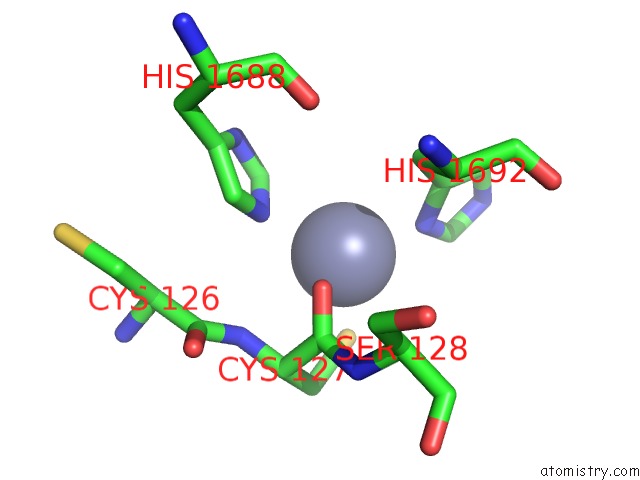
Mono view
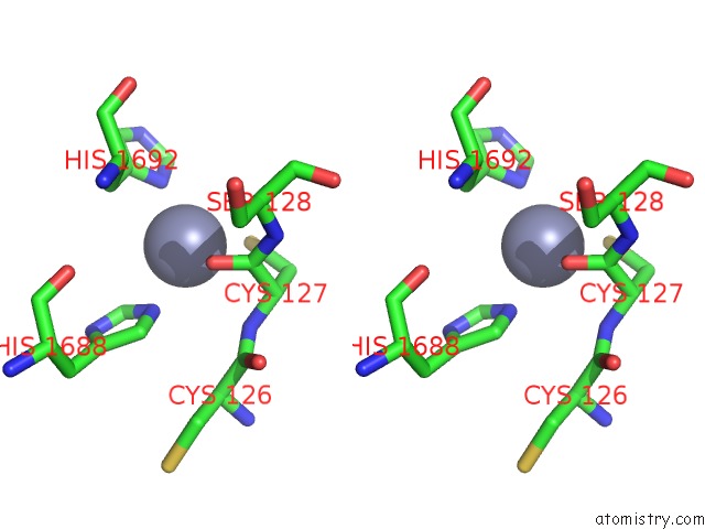
Stereo pair view
|
A full contact list of Zinc with other atoms in the Zn binding
site number 6 of X-Ray Structure of Pedv Papain-Like Protease 2 Bound to Ub-Pa within 5.0Å range:
probe
|
atom
|
residue
|
distance (Å)
|
B
|
Occ
|
E:Zn2002
b:59.2
occ:1.00
|
O
|
K:CYS127
|
2.3
|
70.1
|
1.0
|
ND1
|
E:HIS1692
|
2.3
|
49.5
|
1.0
|
SG
|
K:CYS127
|
2.5
|
63.1
|
1.0
|
NE2
|
E:HIS1688
|
2.5
|
68.0
|
1.0
|
C
|
K:CYS127
|
3.0
|
69.7
|
1.0
|
CE1
|
E:HIS1688
|
3.0
|
68.5
|
1.0
|
CG
|
E:HIS1692
|
3.3
|
52.3
|
1.0
|
CE1
|
E:HIS1692
|
3.3
|
55.3
|
1.0
|
CB
|
E:HIS1692
|
3.5
|
57.0
|
1.0
|
N
|
K:CYS127
|
3.5
|
85.2
|
1.0
|
CA
|
K:CYS127
|
3.5
|
78.6
|
1.0
|
CB
|
K:CYS127
|
3.6
|
64.0
|
1.0
|
CD2
|
E:HIS1688
|
3.7
|
71.8
|
1.0
|
N
|
K:SER128
|
4.0
|
71.1
|
1.0
|
ND1
|
E:HIS1688
|
4.2
|
61.9
|
1.0
|
NE2
|
E:HIS1692
|
4.4
|
53.8
|
1.0
|
CD2
|
E:HIS1692
|
4.4
|
48.3
|
1.0
|
CA
|
E:HIS1692
|
4.4
|
55.9
|
1.0
|
CA
|
K:SER128
|
4.4
|
63.2
|
1.0
|
C
|
K:CYS126
|
4.5
|
81.0
|
1.0
|
CG
|
E:HIS1688
|
4.6
|
73.1
|
1.0
|
O
|
E:HIS1688
|
4.9
|
67.4
|
1.0
|
|
Zinc binding site 7 out
of 14 in 7mc9
Go back to
Zinc Binding Sites List in 7mc9
Zinc binding site 7 out
of 14 in the X-Ray Structure of Pedv Papain-Like Protease 2 Bound to Ub-Pa
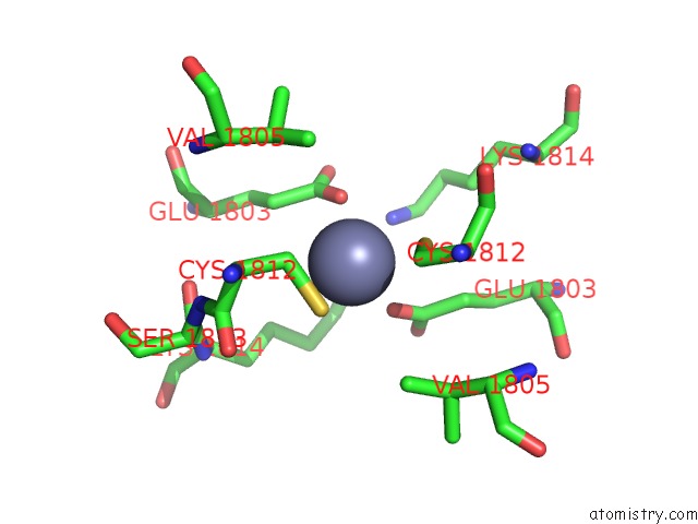
Mono view
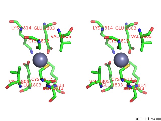
Stereo pair view
|
A full contact list of Zinc with other atoms in the Zn binding
site number 7 of X-Ray Structure of Pedv Papain-Like Protease 2 Bound to Ub-Pa within 5.0Å range:
probe
|
atom
|
residue
|
distance (Å)
|
B
|
Occ
|
E:Zn2003
b:96.2
occ:1.00
|
SG
|
G:CYS1812
|
2.6
|
85.8
|
1.0
|
OE2
|
E:GLU1803
|
2.6
|
65.1
|
1.0
|
OE2
|
G:GLU1803
|
2.8
|
66.3
|
1.0
|
SG
|
E:CYS1812
|
2.9
|
97.3
|
1.0
|
CB
|
E:CYS1812
|
3.3
|
55.4
|
1.0
|
CB
|
G:CYS1812
|
3.5
|
57.6
|
1.0
|
CD
|
E:GLU1803
|
3.6
|
65.6
|
1.0
|
CD
|
G:GLU1803
|
3.9
|
64.8
|
1.0
|
CG
|
E:GLU1803
|
4.0
|
50.0
|
1.0
|
NZ
|
G:LYS1814
|
4.4
|
76.0
|
1.0
|
CG2
|
G:VAL1805
|
4.5
|
49.0
|
1.0
|
CG
|
G:GLU1803
|
4.5
|
67.2
|
1.0
|
CA
|
E:CYS1812
|
4.6
|
48.6
|
1.0
|
OE1
|
E:GLU1803
|
4.6
|
61.2
|
1.0
|
CG2
|
E:VAL1805
|
4.7
|
45.9
|
1.0
|
C
|
E:CYS1812
|
4.8
|
50.7
|
1.0
|
NZ
|
E:LYS1814
|
4.8
|
61.7
|
1.0
|
CB
|
E:VAL1805
|
4.8
|
42.4
|
1.0
|
OE1
|
G:GLU1803
|
4.9
|
63.3
|
1.0
|
CA
|
G:CYS1812
|
5.0
|
49.6
|
1.0
|
N
|
E:SER1813
|
5.0
|
50.5
|
1.0
|
|
Zinc binding site 8 out
of 14 in 7mc9
Go back to
Zinc Binding Sites List in 7mc9
Zinc binding site 8 out
of 14 in the X-Ray Structure of Pedv Papain-Like Protease 2 Bound to Ub-Pa
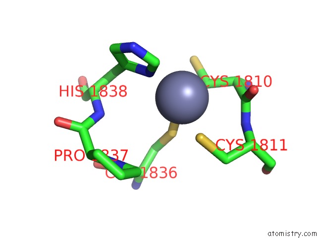
Mono view
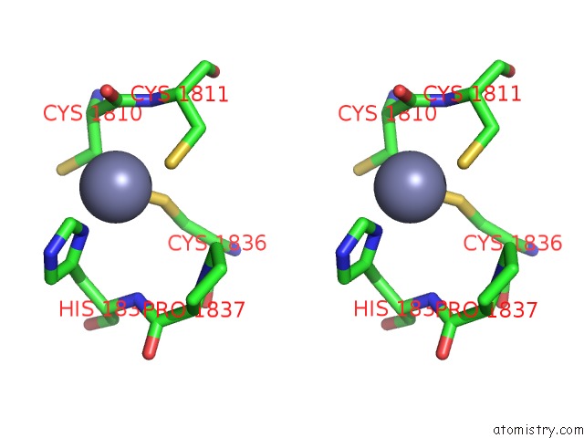
Stereo pair view
|
A full contact list of Zinc with other atoms in the Zn binding
site number 8 of X-Ray Structure of Pedv Papain-Like Protease 2 Bound to Ub-Pa within 5.0Å range:
probe
|
atom
|
residue
|
distance (Å)
|
B
|
Occ
|
G:Zn2001
b:82.4
occ:1.00
|
CB
|
G:CYS1810
|
2.1
|
49.6
|
1.0
|
SG
|
G:CYS1836
|
2.1
|
38.3
|
1.0
|
ND1
|
G:HIS1838
|
2.2
|
76.0
|
1.0
|
SG
|
G:CYS1811
|
2.8
|
54.7
|
1.0
|
CE1
|
G:HIS1838
|
3.0
|
77.0
|
1.0
|
SG
|
G:CYS1810
|
3.2
|
97.2
|
1.0
|
CA
|
G:CYS1810
|
3.2
|
55.1
|
1.0
|
C
|
G:CYS1810
|
3.3
|
64.0
|
1.0
|
CG
|
G:HIS1838
|
3.4
|
71.0
|
1.0
|
N
|
G:CYS1811
|
3.6
|
59.7
|
1.0
|
CB
|
G:CYS1836
|
3.8
|
55.1
|
1.0
|
O
|
G:CYS1810
|
3.8
|
66.5
|
1.0
|
CB
|
G:HIS1838
|
3.9
|
62.0
|
1.0
|
CB
|
G:CYS1811
|
4.0
|
45.1
|
1.0
|
NE2
|
G:HIS1838
|
4.2
|
96.3
|
1.0
|
CA
|
G:CYS1811
|
4.3
|
38.2
|
1.0
|
N
|
G:CYS1810
|
4.4
|
56.5
|
1.0
|
N
|
G:HIS1838
|
4.4
|
60.4
|
1.0
|
CD2
|
G:HIS1838
|
4.4
|
94.1
|
1.0
|
CD
|
G:PRO1837
|
4.6
|
60.5
|
1.0
|
CA
|
G:HIS1838
|
4.8
|
63.5
|
1.0
|
N
|
G:PRO1837
|
4.8
|
65.4
|
1.0
|
CA
|
G:CYS1836
|
4.8
|
61.6
|
1.0
|
|
Zinc binding site 9 out
of 14 in 7mc9
Go back to
Zinc Binding Sites List in 7mc9
Zinc binding site 9 out
of 14 in the X-Ray Structure of Pedv Papain-Like Protease 2 Bound to Ub-Pa
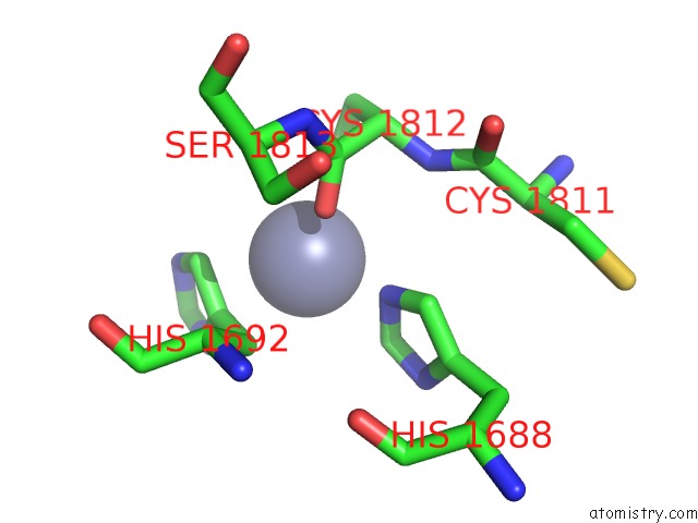
Mono view
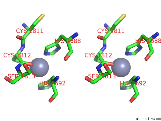
Stereo pair view
|
A full contact list of Zinc with other atoms in the Zn binding
site number 9 of X-Ray Structure of Pedv Papain-Like Protease 2 Bound to Ub-Pa within 5.0Å range:
probe
|
atom
|
residue
|
distance (Å)
|
B
|
Occ
|
G:Zn2002
b:47.1
occ:1.00
|
NE2
|
G:HIS1688
|
2.3
|
48.0
|
1.0
|
O
|
I:CYS1812
|
2.4
|
42.3
|
1.0
|
CG
|
G:HIS1692
|
2.6
|
54.7
|
1.0
|
CD2
|
G:HIS1692
|
2.6
|
57.4
|
1.0
|
SG
|
I:CYS1812
|
2.6
|
60.9
|
1.0
|
CE1
|
G:HIS1688
|
2.8
|
47.5
|
1.0
|
CB
|
G:HIS1692
|
3.1
|
47.7
|
1.0
|
ND1
|
G:HIS1692
|
3.1
|
51.9
|
1.0
|
NE2
|
G:HIS1692
|
3.1
|
46.8
|
1.0
|
C
|
I:CYS1812
|
3.3
|
53.5
|
1.0
|
CE1
|
G:HIS1692
|
3.4
|
49.2
|
1.0
|
CD2
|
G:HIS1688
|
3.6
|
61.5
|
1.0
|
CB
|
I:CYS1812
|
3.8
|
57.9
|
1.0
|
N
|
I:CYS1812
|
3.9
|
61.1
|
1.0
|
CA
|
I:CYS1812
|
3.9
|
48.9
|
1.0
|
ND1
|
G:HIS1688
|
4.0
|
46.5
|
1.0
|
CA
|
G:HIS1692
|
4.3
|
48.6
|
1.0
|
N
|
I:SER1813
|
4.4
|
57.8
|
1.0
|
CG
|
G:HIS1688
|
4.4
|
57.2
|
1.0
|
CA
|
I:SER1813
|
4.8
|
43.7
|
1.0
|
C
|
I:CYS1811
|
5.0
|
58.1
|
1.0
|
|
Zinc binding site 10 out
of 14 in 7mc9
Go back to
Zinc Binding Sites List in 7mc9
Zinc binding site 10 out
of 14 in the X-Ray Structure of Pedv Papain-Like Protease 2 Bound to Ub-Pa
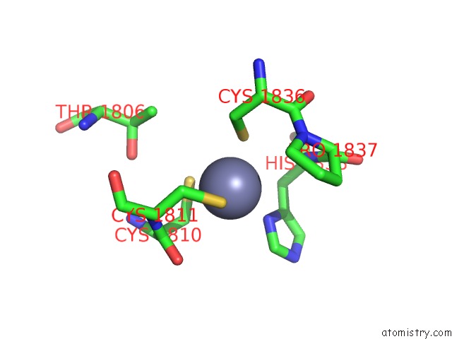
Mono view
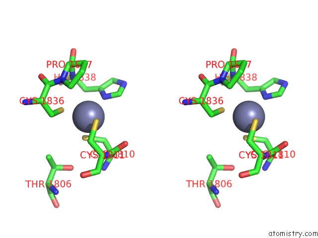
Stereo pair view
|
A full contact list of Zinc with other atoms in the Zn binding
site number 10 of X-Ray Structure of Pedv Papain-Like Protease 2 Bound to Ub-Pa within 5.0Å range:
probe
|
atom
|
residue
|
distance (Å)
|
B
|
Occ
|
I:Zn2001
b:55.1
occ:1.00
|
SG
|
I:CYS1810
|
2.3
|
52.8
|
1.0
|
ND1
|
I:HIS1838
|
2.4
|
59.4
|
1.0
|
SG
|
I:CYS1811
|
2.4
|
77.9
|
1.0
|
SG
|
I:CYS1836
|
2.5
|
48.4
|
1.0
|
CB
|
I:CYS1810
|
3.1
|
52.6
|
1.0
|
CB
|
I:CYS1836
|
3.4
|
54.8
|
1.0
|
CE1
|
I:HIS1838
|
3.4
|
57.6
|
1.0
|
CG
|
I:HIS1838
|
3.4
|
56.5
|
1.0
|
CB
|
I:CYS1811
|
3.6
|
52.8
|
1.0
|
CB
|
I:HIS1838
|
3.6
|
46.7
|
1.0
|
C
|
I:CYS1810
|
3.7
|
61.7
|
1.0
|
N
|
I:CYS1811
|
3.8
|
51.7
|
1.0
|
N
|
I:HIS1838
|
3.8
|
47.0
|
1.0
|
O
|
I:CYS1810
|
4.0
|
63.5
|
1.0
|
CA
|
I:CYS1810
|
4.0
|
55.4
|
1.0
|
CD
|
I:PRO1837
|
4.2
|
52.2
|
1.0
|
N
|
I:PRO1837
|
4.3
|
53.0
|
1.0
|
CA
|
I:CYS1811
|
4.3
|
49.3
|
1.0
|
CA
|
I:HIS1838
|
4.3
|
46.0
|
1.0
|
NE2
|
I:HIS1838
|
4.5
|
51.5
|
1.0
|
CD2
|
I:HIS1838
|
4.5
|
58.2
|
1.0
|
CA
|
I:CYS1836
|
4.5
|
48.2
|
1.0
|
C
|
I:CYS1836
|
4.6
|
54.8
|
1.0
|
C
|
I:PRO1837
|
4.8
|
52.5
|
1.0
|
N
|
I:CYS1810
|
4.9
|
52.1
|
1.0
|
CG2
|
I:THR1806
|
4.9
|
52.5
|
1.0
|
OG1
|
I:THR1806
|
4.9
|
59.2
|
1.0
|
CA
|
I:PRO1837
|
4.9
|
57.7
|
1.0
|
|
Reference:
I.A.Durie,
J.V.Dzimianski,
C.M.Daczkowski,
J.Mcguire,
K.Faaberg,
S.D.Pegan.
Structural Insights Into the Interaction of Papain-Like Protease 2 From the Alphacoronavirus Porcine Epidemic Diarrhea Virus and Ubiquitin Acta Cryst. D V. 77 943 2021.
DOI: 10.1107/S205979832100509X
Page generated: Tue Oct 29 23:12:17 2024
|