Zinc »
PDB 7esi-7f9c »
7f5i »
Zinc in PDB 7f5i: X-Ray Structure of Clostridium Perfringens-Specific Amidase Endolysin
Protein crystallography data
The structure of X-Ray Structure of Clostridium Perfringens-Specific Amidase Endolysin, PDB code: 7f5i
was solved by
S.Kamitori,
E.Tamai,
with X-Ray Crystallography technique. A brief refinement statistics is given in the table below:
| Resolution Low / High (Å) | 18.36 / 1.65 |
| Space group | P 21 21 21 |
| Cell size a, b, c (Å), α, β, γ (°) | 51.01, 52.87, 58.61, 90, 90, 90 |
| R / Rfree (%) | 13.7 / 20.5 |
Other elements in 7f5i:
The structure of X-Ray Structure of Clostridium Perfringens-Specific Amidase Endolysin also contains other interesting chemical elements:
| Sodium | (Na) | 1 atom |
Zinc Binding Sites:
The binding sites of Zinc atom in the X-Ray Structure of Clostridium Perfringens-Specific Amidase Endolysin
(pdb code 7f5i). This binding sites where shown within
5.0 Angstroms radius around Zinc atom.
In total only one binding site of Zinc was determined in the X-Ray Structure of Clostridium Perfringens-Specific Amidase Endolysin, PDB code: 7f5i:
In total only one binding site of Zinc was determined in the X-Ray Structure of Clostridium Perfringens-Specific Amidase Endolysin, PDB code: 7f5i:
Zinc binding site 1 out of 1 in 7f5i
Go back to
Zinc binding site 1 out
of 1 in the X-Ray Structure of Clostridium Perfringens-Specific Amidase Endolysin
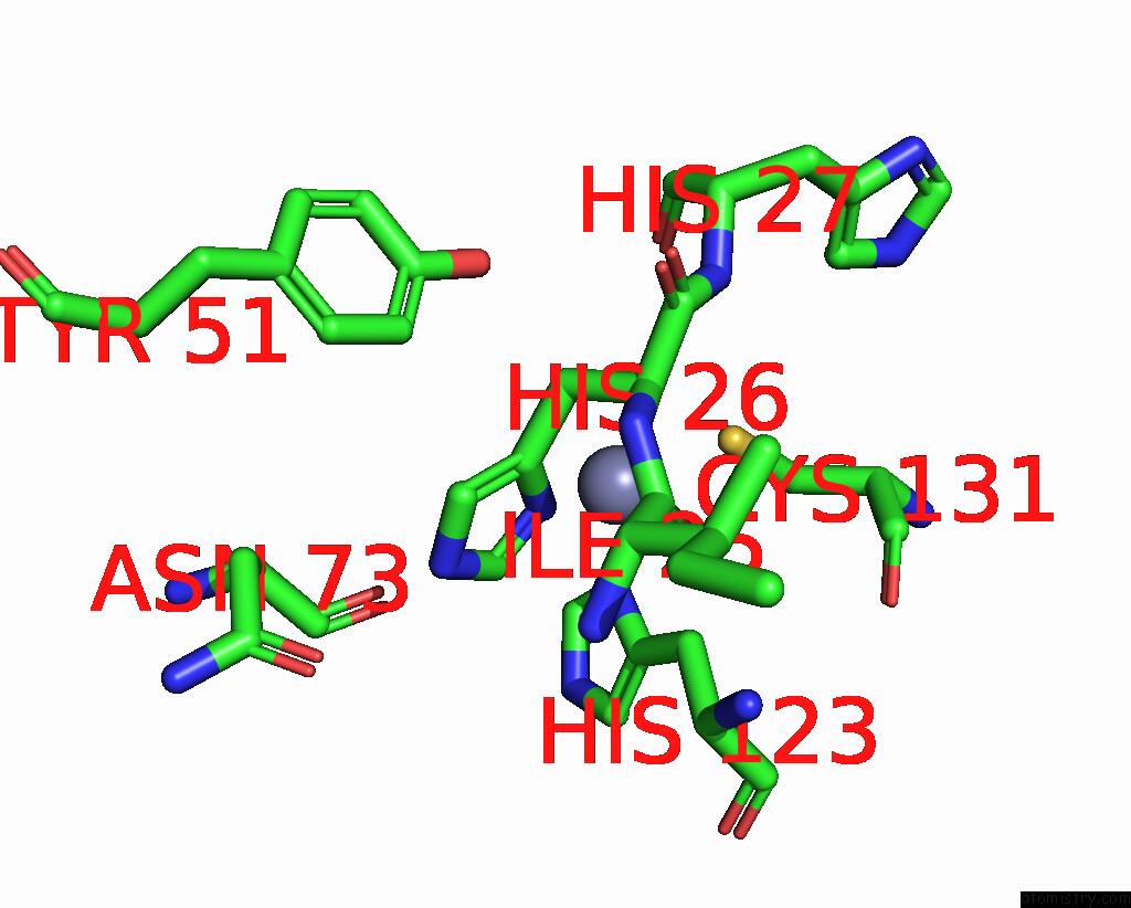
Mono view
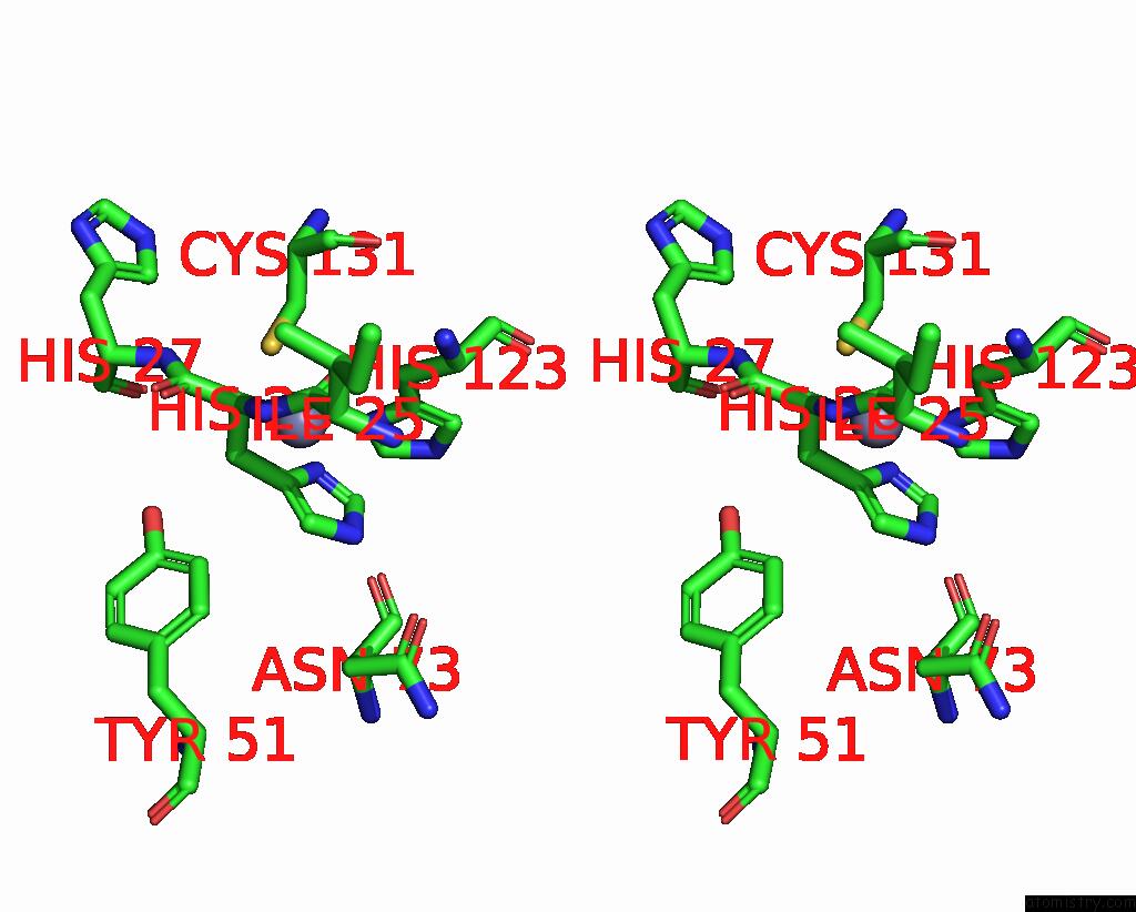
Stereo pair view
Mono view
Stereo pair view
A full contact list of Zinc with other atoms in the Zn binding
site number 1 of X-Ray Structure of Clostridium Perfringens-Specific Amidase Endolysin within 5.0Å range:
|
Reference:
H.Sekiya,
S.Kamitori,
H.Nariya,
R.Matsunami,
E.Tamai.
Structural and Biochemical Characterization of the Clostridium Perfringens-Specific Zn 2+ -Dependent Amidase Endolysin, Psa, Catalytic Domain. Biochem.Biophys.Res.Commun. V. 576 66 2021.
ISSN: ESSN 1090-2104
PubMed: 34482025
DOI: 10.1016/J.BBRC.2021.08.085
Page generated: Tue Oct 29 20:13:01 2024
ISSN: ESSN 1090-2104
PubMed: 34482025
DOI: 10.1016/J.BBRC.2021.08.085
Last articles
Zn in 9MJ5Zn in 9HNW
Zn in 9G0L
Zn in 9FNE
Zn in 9DZN
Zn in 9E0I
Zn in 9D32
Zn in 9DAK
Zn in 8ZXC
Zn in 8ZUF