Zinc »
PDB 7esi-7f9c »
7exv »
Zinc in PDB 7exv: GH127 Beta-L-Arabinofuranosidase HYPBA1 Covalently Complexed with P- Nitrophenyl Beta-L-Arabinofuranoylamide
Enzymatic activity of GH127 Beta-L-Arabinofuranosidase HYPBA1 Covalently Complexed with P- Nitrophenyl Beta-L-Arabinofuranoylamide
All present enzymatic activity of GH127 Beta-L-Arabinofuranosidase HYPBA1 Covalently Complexed with P- Nitrophenyl Beta-L-Arabinofuranoylamide:
3.2.1.185;
3.2.1.185;
Protein crystallography data
The structure of GH127 Beta-L-Arabinofuranosidase HYPBA1 Covalently Complexed with P- Nitrophenyl Beta-L-Arabinofuranoylamide, PDB code: 7exv
was solved by
K.Sawano,
T.Arakawa,
C.Yamada,
K.Fujita,
S.Fushinobu,
with X-Ray Crystallography technique. A brief refinement statistics is given in the table below:
| Resolution Low / High (Å) | 46.14 / 2.60 |
| Space group | P 32 2 1 |
| Cell size a, b, c (Å), α, β, γ (°) | 77.547, 77.547, 253.529, 90, 90, 120 |
| R / Rfree (%) | 25.1 / 31 |
Zinc Binding Sites:
The binding sites of Zinc atom in the GH127 Beta-L-Arabinofuranosidase HYPBA1 Covalently Complexed with P- Nitrophenyl Beta-L-Arabinofuranoylamide
(pdb code 7exv). This binding sites where shown within
5.0 Angstroms radius around Zinc atom.
In total only one binding site of Zinc was determined in the GH127 Beta-L-Arabinofuranosidase HYPBA1 Covalently Complexed with P- Nitrophenyl Beta-L-Arabinofuranoylamide, PDB code: 7exv:
In total only one binding site of Zinc was determined in the GH127 Beta-L-Arabinofuranosidase HYPBA1 Covalently Complexed with P- Nitrophenyl Beta-L-Arabinofuranoylamide, PDB code: 7exv:
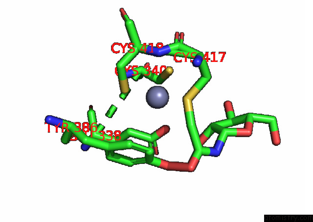
Zinc binding site 1 out of 1 in 7exv
Go back to
Zinc binding site 1 out
of 1 in the GH127 Beta-L-Arabinofuranosidase HYPBA1 Covalently Complexed with P- Nitrophenyl Beta-L-Arabinofuranoylamide
Mono view

Stereo pair view
Mono view
Stereo pair view
A full contact list of Zinc with other atoms in the Zn binding
site number 1 of GH127 Beta-L-Arabinofuranosidase HYPBA1 Covalently Complexed with P- Nitrophenyl Beta-L-Arabinofuranoylamide within 5.0Å range:
|
Reference:
S.Maruyama,
K.Sawano,
S.Amaki,
T.Suzuki,
S.Narita,
K.Kimura,
T.Arakawa,
C.Yamada,
Y.Ito,
N.Dohmae,
K.Fujita,
A.Ishiwata,
S.Fushinobu.
Substrate Complex Structure, Active Site Labeling and Catalytic Role of the Zinc Ion in Cysteine Glycosidase. Glycobiology 2021.
ISSN: ESSN 1460-2423
PubMed: 34735571
DOI: 10.1093/GLYCOB/CWAB103
Page generated: Tue Oct 29 20:04:07 2024
ISSN: ESSN 1460-2423
PubMed: 34735571
DOI: 10.1093/GLYCOB/CWAB103
Last articles
Zn in 9MJ5Zn in 9HNW
Zn in 9G0L
Zn in 9FNE
Zn in 9DZN
Zn in 9E0I
Zn in 9D32
Zn in 9DAK
Zn in 8ZXC
Zn in 8ZUF