Zinc »
PDB 7doy-7dws »
7drj »
Zinc in PDB 7drj: Crystal Structure of Phosphatidylglycerol Phosphate Synthase in Complex with Phosphatidylglycerol Phosphate
Enzymatic activity of Crystal Structure of Phosphatidylglycerol Phosphate Synthase in Complex with Phosphatidylglycerol Phosphate
All present enzymatic activity of Crystal Structure of Phosphatidylglycerol Phosphate Synthase in Complex with Phosphatidylglycerol Phosphate:
2.7.8.5;
2.7.8.5;
Protein crystallography data
The structure of Crystal Structure of Phosphatidylglycerol Phosphate Synthase in Complex with Phosphatidylglycerol Phosphate, PDB code: 7drj
was solved by
B.W.Yang,
Z.F.Liu,
with X-Ray Crystallography technique. A brief refinement statistics is given in the table below:
| Resolution Low / High (Å) | 31.46 / 2.50 |
| Space group | C 1 2 1 |
| Cell size a, b, c (Å), α, β, γ (°) | 127.54, 59.376, 72.985, 90, 106.18, 90 |
| R / Rfree (%) | 21.2 / 24.9 |
Zinc Binding Sites:
The binding sites of Zinc atom in the Crystal Structure of Phosphatidylglycerol Phosphate Synthase in Complex with Phosphatidylglycerol Phosphate
(pdb code 7drj). This binding sites where shown within
5.0 Angstroms radius around Zinc atom.
In total 4 binding sites of Zinc where determined in the Crystal Structure of Phosphatidylglycerol Phosphate Synthase in Complex with Phosphatidylglycerol Phosphate, PDB code: 7drj:
Jump to Zinc binding site number: 1; 2; 3; 4;
In total 4 binding sites of Zinc where determined in the Crystal Structure of Phosphatidylglycerol Phosphate Synthase in Complex with Phosphatidylglycerol Phosphate, PDB code: 7drj:
Jump to Zinc binding site number: 1; 2; 3; 4;
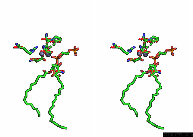
Zinc binding site 1 out of 4 in 7drj
Go back to
Zinc binding site 1 out
of 4 in the Crystal Structure of Phosphatidylglycerol Phosphate Synthase in Complex with Phosphatidylglycerol Phosphate

Mono view

Stereo pair view
Mono view
Stereo pair view
A full contact list of Zinc with other atoms in the Zn binding
site number 1 of Crystal Structure of Phosphatidylglycerol Phosphate Synthase in Complex with Phosphatidylglycerol Phosphate within 5.0Å range:
|
Zinc binding site 2 out of 4 in 7drj
Go back to
Zinc binding site 2 out
of 4 in the Crystal Structure of Phosphatidylglycerol Phosphate Synthase in Complex with Phosphatidylglycerol Phosphate

Mono view

Stereo pair view
Mono view
Stereo pair view
A full contact list of Zinc with other atoms in the Zn binding
site number 2 of Crystal Structure of Phosphatidylglycerol Phosphate Synthase in Complex with Phosphatidylglycerol Phosphate within 5.0Å range:
|
Zinc binding site 3 out of 4 in 7drj
Go back to
Zinc binding site 3 out
of 4 in the Crystal Structure of Phosphatidylglycerol Phosphate Synthase in Complex with Phosphatidylglycerol Phosphate

Mono view
Stereo pair view
Mono view
Stereo pair view
A full contact list of Zinc with other atoms in the Zn binding
site number 3 of Crystal Structure of Phosphatidylglycerol Phosphate Synthase in Complex with Phosphatidylglycerol Phosphate within 5.0Å range:
|
Zinc binding site 4 out of 4 in 7drj
Go back to
Zinc binding site 4 out
of 4 in the Crystal Structure of Phosphatidylglycerol Phosphate Synthase in Complex with Phosphatidylglycerol Phosphate

Mono view

Stereo pair view
Mono view
Stereo pair view
A full contact list of Zinc with other atoms in the Zn binding
site number 4 of Crystal Structure of Phosphatidylglycerol Phosphate Synthase in Complex with Phosphatidylglycerol Phosphate within 5.0Å range:
|
Reference:
B.Yang,
H.Yao,
D.Li,
Z.Liu.
The Phosphatidylglycerol Phosphate Synthase Pgsa Utilizes A Trifurcated Amphipathic Cavity For Catalysis at the Membrane-Cytosol Interface Curr Res Struct Biol 2021.
ISSN: ESSN 2665-928X
DOI: 10.1016/J.CRSTBI.2021.11.005
Page generated: Tue Oct 29 19:14:39 2024
ISSN: ESSN 2665-928X
DOI: 10.1016/J.CRSTBI.2021.11.005
Last articles
Al in 7VPJAl in 7TTT
Al in 7U1Y
Al in 7TTN
Al in 7TRG
Al in 7T3C
Al in 7T3B
Al in 7TQY
Al in 7T22
Al in 7T21