Zinc »
PDB 7crq-7d4q »
7d21 »
Zinc in PDB 7d21: Crystal Structure of Ixodes Scapularis Glutaminyl Cyclase with Two Zn Ions Bound to the Active Site
Enzymatic activity of Crystal Structure of Ixodes Scapularis Glutaminyl Cyclase with Two Zn Ions Bound to the Active Site
All present enzymatic activity of Crystal Structure of Ixodes Scapularis Glutaminyl Cyclase with Two Zn Ions Bound to the Active Site:
2.3.2.5;
2.3.2.5;
Protein crystallography data
The structure of Crystal Structure of Ixodes Scapularis Glutaminyl Cyclase with Two Zn Ions Bound to the Active Site, PDB code: 7d21
was solved by
K.-F.Huang,
J.-S.Huang,
M.-L.Wu,
W.-L.Hsieh,
A.H.-J.Wang,
with X-Ray Crystallography technique. A brief refinement statistics is given in the table below:
| Resolution Low / High (Å) | 27.84 / 1.97 |
| Space group | P 21 21 21 |
| Cell size a, b, c (Å), α, β, γ (°) | 55.646, 70.932, 80.556, 90, 90, 90 |
| R / Rfree (%) | 18.9 / 22.2 |
Zinc Binding Sites:
The binding sites of Zinc atom in the Crystal Structure of Ixodes Scapularis Glutaminyl Cyclase with Two Zn Ions Bound to the Active Site
(pdb code 7d21). This binding sites where shown within
5.0 Angstroms radius around Zinc atom.
In total 5 binding sites of Zinc where determined in the Crystal Structure of Ixodes Scapularis Glutaminyl Cyclase with Two Zn Ions Bound to the Active Site, PDB code: 7d21:
Jump to Zinc binding site number: 1; 2; 3; 4; 5;
In total 5 binding sites of Zinc where determined in the Crystal Structure of Ixodes Scapularis Glutaminyl Cyclase with Two Zn Ions Bound to the Active Site, PDB code: 7d21:
Jump to Zinc binding site number: 1; 2; 3; 4; 5;
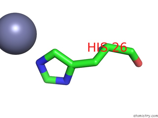
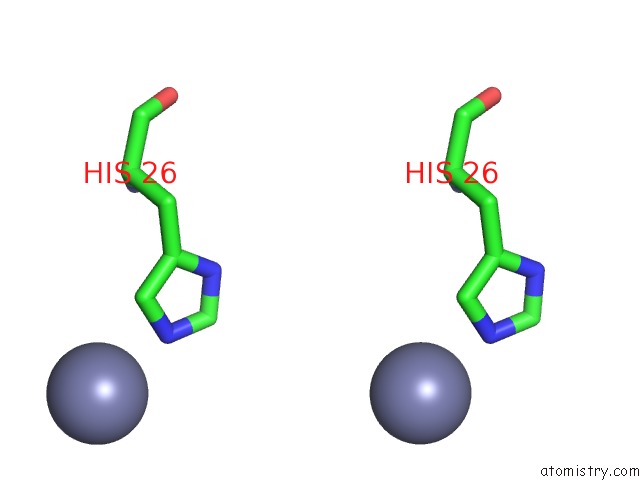
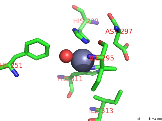
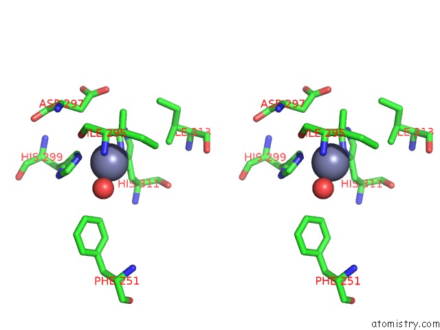
Zinc binding site 1 out of 5 in 7d21
Go back to
Zinc binding site 1 out
of 5 in the Crystal Structure of Ixodes Scapularis Glutaminyl Cyclase with Two Zn Ions Bound to the Active Site
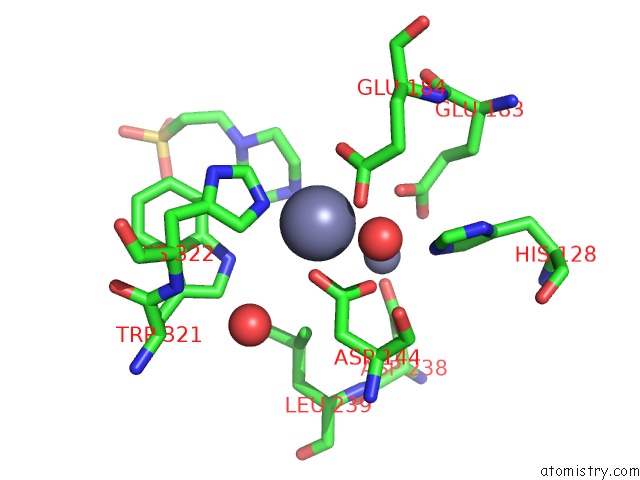
Mono view
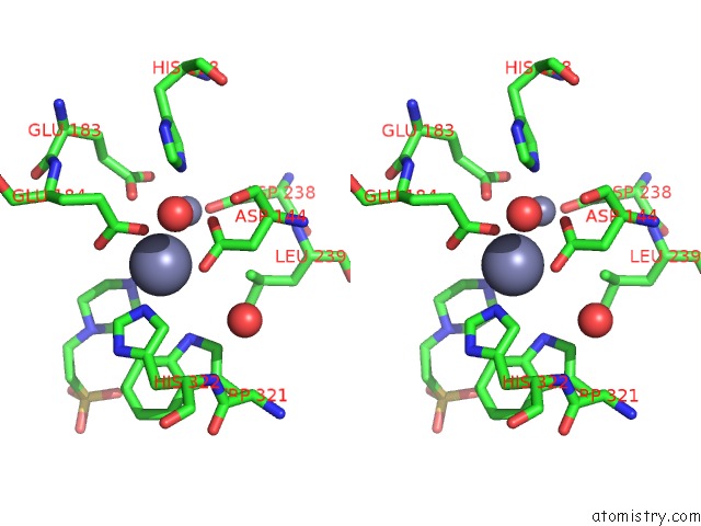
Stereo pair view
Mono view
Stereo pair view
A full contact list of Zinc with other atoms in the Zn binding
site number 1 of Crystal Structure of Ixodes Scapularis Glutaminyl Cyclase with Two Zn Ions Bound to the Active Site within 5.0Å range:
|
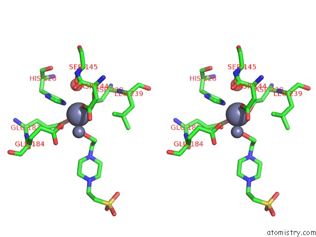
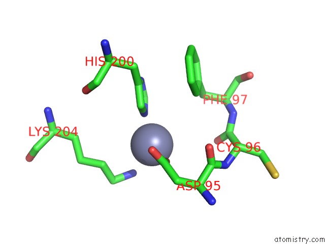
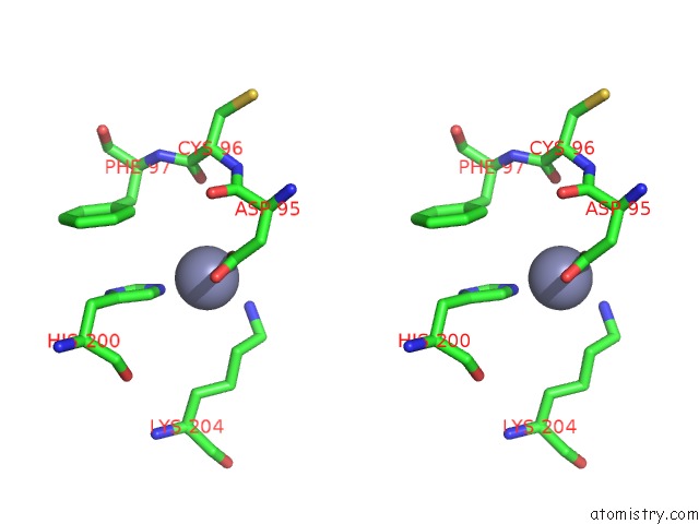
Zinc binding site 2 out of 5 in 7d21
Go back to
Zinc binding site 2 out
of 5 in the Crystal Structure of Ixodes Scapularis Glutaminyl Cyclase with Two Zn Ions Bound to the Active Site
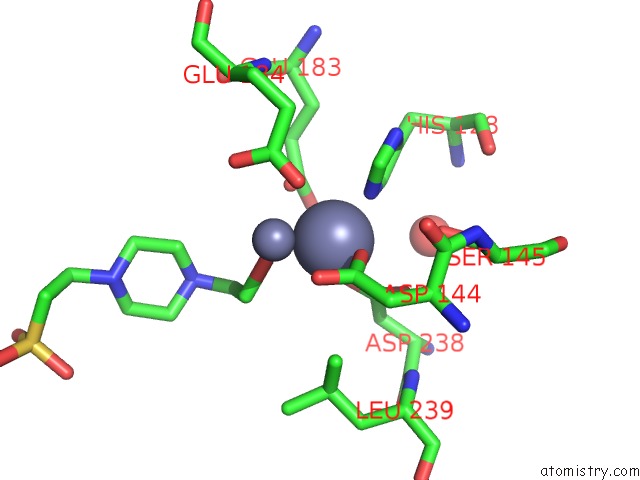
Mono view
Stereo pair view
Mono view
Stereo pair view
A full contact list of Zinc with other atoms in the Zn binding
site number 2 of Crystal Structure of Ixodes Scapularis Glutaminyl Cyclase with Two Zn Ions Bound to the Active Site within 5.0Å range:
|
Zinc binding site 3 out of 5 in 7d21
Go back to
Zinc binding site 3 out
of 5 in the Crystal Structure of Ixodes Scapularis Glutaminyl Cyclase with Two Zn Ions Bound to the Active Site
Mono view
Stereo pair view
Mono view
Stereo pair view
A full contact list of Zinc with other atoms in the Zn binding
site number 3 of Crystal Structure of Ixodes Scapularis Glutaminyl Cyclase with Two Zn Ions Bound to the Active Site within 5.0Å range:
|
Zinc binding site 4 out of 5 in 7d21
Go back to
Zinc binding site 4 out
of 5 in the Crystal Structure of Ixodes Scapularis Glutaminyl Cyclase with Two Zn Ions Bound to the Active Site
Mono view
Stereo pair view
Mono view
Stereo pair view
A full contact list of Zinc with other atoms in the Zn binding
site number 4 of Crystal Structure of Ixodes Scapularis Glutaminyl Cyclase with Two Zn Ions Bound to the Active Site within 5.0Å range:
|
Zinc binding site 5 out of 5 in 7d21
Go back to
Zinc binding site 5 out
of 5 in the Crystal Structure of Ixodes Scapularis Glutaminyl Cyclase with Two Zn Ions Bound to the Active Site
Mono view
Stereo pair view
Mono view
Stereo pair view
A full contact list of Zinc with other atoms in the Zn binding
site number 5 of Crystal Structure of Ixodes Scapularis Glutaminyl Cyclase with Two Zn Ions Bound to the Active Site within 5.0Å range:
|
Reference:
K.F.Huang,
J.S.Huang,
M.L.Wu,
W.L.Hsieh,
K.C.Hsu,
H.L.Hsu,
T.P.Ko,
A.H-J Wang.
A Unique Carboxylic-Acid Hydrogen-Bond Network (Cahbn) Confers Glutaminyl Cyclase Activity on M28 Family Enzymes. J.Mol.Biol. 66960 2021.
ISSN: ESSN 1089-8638
PubMed: 33774034
DOI: 10.1016/J.JMB.2021.166960
Page generated: Tue Oct 29 18:36:22 2024
ISSN: ESSN 1089-8638
PubMed: 33774034
DOI: 10.1016/J.JMB.2021.166960
Last articles
Zn in 9MJ5Zn in 9HNW
Zn in 9G0L
Zn in 9FNE
Zn in 9DZN
Zn in 9E0I
Zn in 9D32
Zn in 9DAK
Zn in 8ZXC
Zn in 8ZUF