Zinc »
PDB 7a91-7aes »
7abw »
Zinc in PDB 7abw: Crystal Structure of Siderophore Reductase Foxb
Protein crystallography data
The structure of Crystal Structure of Siderophore Reductase Foxb, PDB code: 7abw
was solved by
I.Josts,
H.Tidow,
with X-Ray Crystallography technique. A brief refinement statistics is given in the table below:
| Resolution Low / High (Å) | 46.36 / 3.35 |
| Space group | P 21 21 2 |
| Cell size a, b, c (Å), α, β, γ (°) | 237.065, 114.321, 64.382, 90, 90, 90 |
| R / Rfree (%) | 26.5 / 29.9 |
Other elements in 7abw:
The structure of Crystal Structure of Siderophore Reductase Foxb also contains other interesting chemical elements:
| Iron | (Fe) | 4 atoms |
Zinc Binding Sites:
The binding sites of Zinc atom in the Crystal Structure of Siderophore Reductase Foxb
(pdb code 7abw). This binding sites where shown within
5.0 Angstroms radius around Zinc atom.
In total 3 binding sites of Zinc where determined in the Crystal Structure of Siderophore Reductase Foxb, PDB code: 7abw:
Jump to Zinc binding site number: 1; 2; 3;
In total 3 binding sites of Zinc where determined in the Crystal Structure of Siderophore Reductase Foxb, PDB code: 7abw:
Jump to Zinc binding site number: 1; 2; 3;
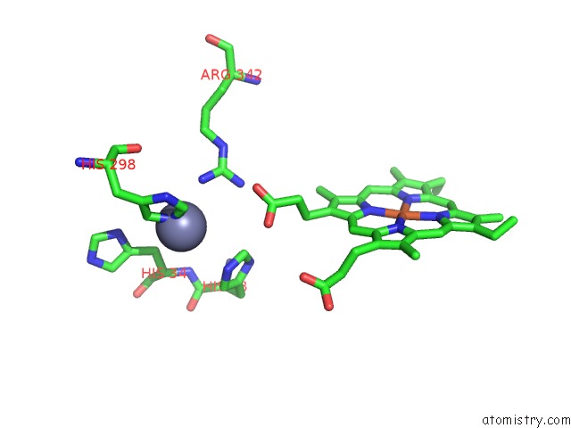
Zinc binding site 1 out of 3 in 7abw
Go back to
Zinc binding site 1 out
of 3 in the Crystal Structure of Siderophore Reductase Foxb
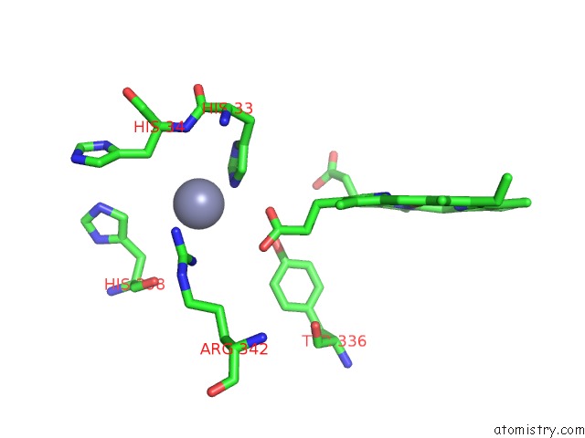
Mono view
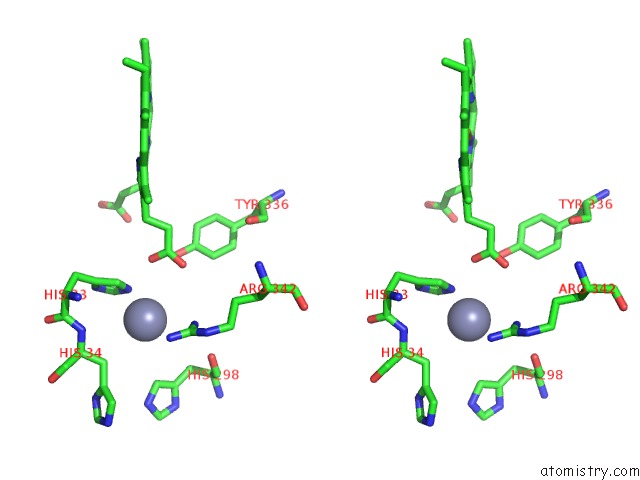
Stereo pair view
Mono view
Stereo pair view
A full contact list of Zinc with other atoms in the Zn binding
site number 1 of Crystal Structure of Siderophore Reductase Foxb within 5.0Å range:
|
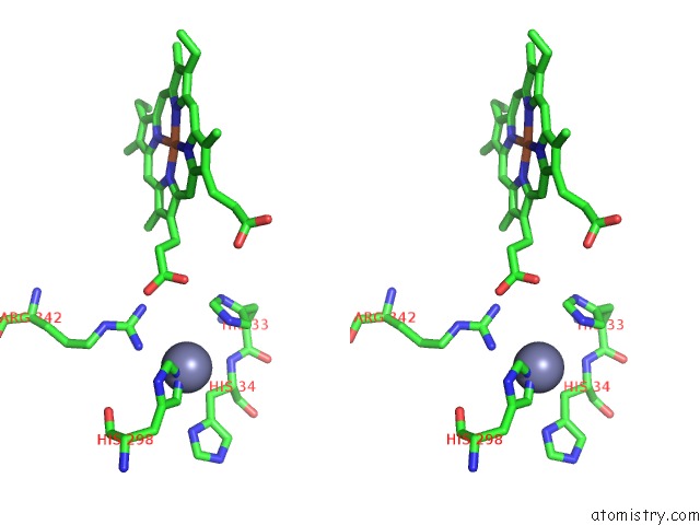
Zinc binding site 2 out of 3 in 7abw
Go back to
Zinc binding site 2 out
of 3 in the Crystal Structure of Siderophore Reductase Foxb
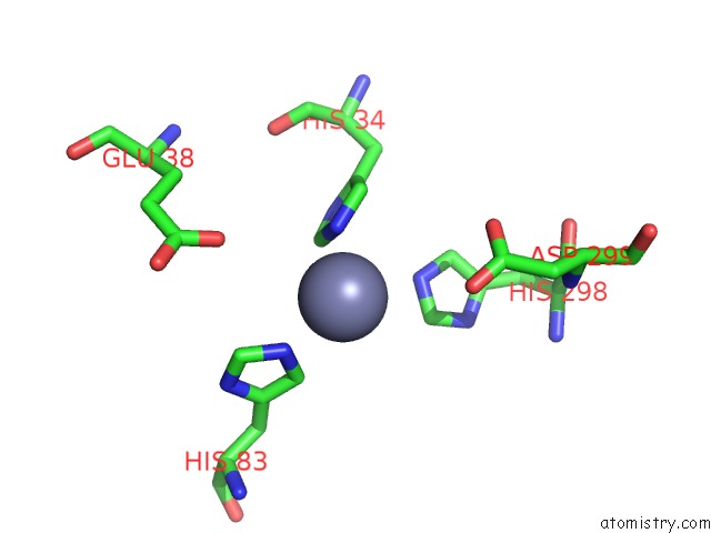
Mono view
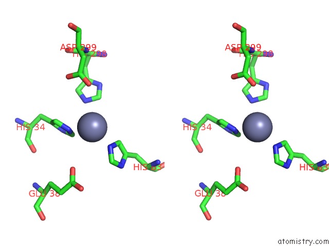
Stereo pair view
Mono view
Stereo pair view
A full contact list of Zinc with other atoms in the Zn binding
site number 2 of Crystal Structure of Siderophore Reductase Foxb within 5.0Å range:
|
Zinc binding site 3 out of 3 in 7abw
Go back to
Zinc binding site 3 out
of 3 in the Crystal Structure of Siderophore Reductase Foxb
Mono view
Stereo pair view
Mono view
Stereo pair view
A full contact list of Zinc with other atoms in the Zn binding
site number 3 of Crystal Structure of Siderophore Reductase Foxb within 5.0Å range:
|
Reference:
I.Josts,
K.Veith,
V.Normant,
I.J.Schalk,
H.Tidow.
Structural Insights Into A Novel Family of Integral Membrane Siderophore Reductases. Proc.Natl.Acad.Sci.Usa V. 118 2021.
ISSN: ESSN 1091-6490
PubMed: 34417315
DOI: 10.1073/PNAS.2101952118
Page generated: Tue Oct 29 16:36:47 2024
ISSN: ESSN 1091-6490
PubMed: 34417315
DOI: 10.1073/PNAS.2101952118
Last articles
Zn in 9MJ5Zn in 9HNW
Zn in 9G0L
Zn in 9FNE
Zn in 9DZN
Zn in 9E0I
Zn in 9D32
Zn in 9DAK
Zn in 8ZXC
Zn in 8ZUF