Zinc »
PDB 6yo4-6yzo »
6yyf »
Zinc in PDB 6yyf: Crystal Structure of 5-Chloroindoline-2,3-Dione Covalently Bound to the pH Domain of Bruton'S Tyrosine Kinase Mutant R28C
Enzymatic activity of Crystal Structure of 5-Chloroindoline-2,3-Dione Covalently Bound to the pH Domain of Bruton'S Tyrosine Kinase Mutant R28C
All present enzymatic activity of Crystal Structure of 5-Chloroindoline-2,3-Dione Covalently Bound to the pH Domain of Bruton'S Tyrosine Kinase Mutant R28C:
2.7.10.2;
2.7.10.2;
Protein crystallography data
The structure of Crystal Structure of 5-Chloroindoline-2,3-Dione Covalently Bound to the pH Domain of Bruton'S Tyrosine Kinase Mutant R28C, PDB code: 6yyf
was solved by
P.Brear,
J.Wagstaff,
M.Hyvonen,
with X-Ray Crystallography technique. A brief refinement statistics is given in the table below:
| Resolution Low / High (Å) | 36.74 / 1.93 |
| Space group | P 1 21 1 |
| Cell size a, b, c (Å), α, β, γ (°) | 46.9, 60.29, 57.4, 90, 98.93, 90 |
| R / Rfree (%) | 19 / 24.9 |
Other elements in 6yyf:
The structure of Crystal Structure of 5-Chloroindoline-2,3-Dione Covalently Bound to the pH Domain of Bruton'S Tyrosine Kinase Mutant R28C also contains other interesting chemical elements:
| Chlorine | (Cl) | 2 atoms |
| Magnesium | (Mg) | 2 atoms |
Zinc Binding Sites:
The binding sites of Zinc atom in the Crystal Structure of 5-Chloroindoline-2,3-Dione Covalently Bound to the pH Domain of Bruton'S Tyrosine Kinase Mutant R28C
(pdb code 6yyf). This binding sites where shown within
5.0 Angstroms radius around Zinc atom.
In total 2 binding sites of Zinc where determined in the Crystal Structure of 5-Chloroindoline-2,3-Dione Covalently Bound to the pH Domain of Bruton'S Tyrosine Kinase Mutant R28C, PDB code: 6yyf:
Jump to Zinc binding site number: 1; 2;
In total 2 binding sites of Zinc where determined in the Crystal Structure of 5-Chloroindoline-2,3-Dione Covalently Bound to the pH Domain of Bruton'S Tyrosine Kinase Mutant R28C, PDB code: 6yyf:
Jump to Zinc binding site number: 1; 2;
Zinc binding site 1 out of 2 in 6yyf
Go back to
Zinc binding site 1 out
of 2 in the Crystal Structure of 5-Chloroindoline-2,3-Dione Covalently Bound to the pH Domain of Bruton'S Tyrosine Kinase Mutant R28C

Mono view

Stereo pair view
Mono view
Stereo pair view
A full contact list of Zinc with other atoms in the Zn binding
site number 1 of Crystal Structure of 5-Chloroindoline-2,3-Dione Covalently Bound to the pH Domain of Bruton'S Tyrosine Kinase Mutant R28C within 5.0Å range:
|
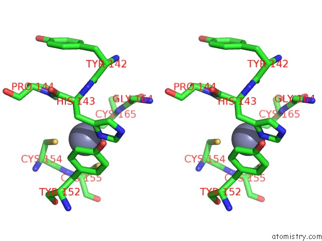
Zinc binding site 2 out of 2 in 6yyf
Go back to
Zinc binding site 2 out
of 2 in the Crystal Structure of 5-Chloroindoline-2,3-Dione Covalently Bound to the pH Domain of Bruton'S Tyrosine Kinase Mutant R28C

Mono view
Stereo pair view
Mono view
Stereo pair view
A full contact list of Zinc with other atoms in the Zn binding
site number 2 of Crystal Structure of 5-Chloroindoline-2,3-Dione Covalently Bound to the pH Domain of Bruton'S Tyrosine Kinase Mutant R28C within 5.0Å range:
|
Reference:
P.Brear,
G.Fischer,
M.May,
T.Pantelejevs,
R.Mathieu,
M.Rossmann,
J.Wagstaff,
B.Blaszczyk,
M.Hyvonen.
Optimising Crystallographic Systems For Structure-Guided Drug Discovery To Be Published.
Page generated: Tue Oct 29 15:17:02 2024
Last articles
Zn in 9MJ5Zn in 9HNW
Zn in 9G0L
Zn in 9FNE
Zn in 9DZN
Zn in 9E0I
Zn in 9D32
Zn in 9DAK
Zn in 8ZXC
Zn in 8ZUF