Zinc »
PDB 6yhq-6yo7 »
6yhr »
Zinc in PDB 6yhr: Crystal Structure of Werner Syndrome Helicase
Enzymatic activity of Crystal Structure of Werner Syndrome Helicase
All present enzymatic activity of Crystal Structure of Werner Syndrome Helicase:
3.6.4.12;
3.6.4.12;
Protein crystallography data
The structure of Crystal Structure of Werner Syndrome Helicase, PDB code: 6yhr
was solved by
J.A.Newman,
A.E.Gavard,
P.Savitsky,
F.Von Delft,
C.H.Arrowsmith,
A.Edwards,
C.Bountra,
O.Gileadi,
with X-Ray Crystallography technique. A brief refinement statistics is given in the table below:
| Resolution Low / High (Å) | 69.12 / 2.20 |
| Space group | P 21 21 21 |
| Cell size a, b, c (Å), α, β, γ (°) | 54.595, 90.627, 138.233, 90.00, 90.00, 90.00 |
| R / Rfree (%) | 19.7 / 23.6 |
Zinc Binding Sites:
The binding sites of Zinc atom in the Crystal Structure of Werner Syndrome Helicase
(pdb code 6yhr). This binding sites where shown within
5.0 Angstroms radius around Zinc atom.
In total 2 binding sites of Zinc where determined in the Crystal Structure of Werner Syndrome Helicase, PDB code: 6yhr:
Jump to Zinc binding site number: 1; 2;
In total 2 binding sites of Zinc where determined in the Crystal Structure of Werner Syndrome Helicase, PDB code: 6yhr:
Jump to Zinc binding site number: 1; 2;
Zinc binding site 1 out of 2 in 6yhr
Go back to
Zinc binding site 1 out
of 2 in the Crystal Structure of Werner Syndrome Helicase
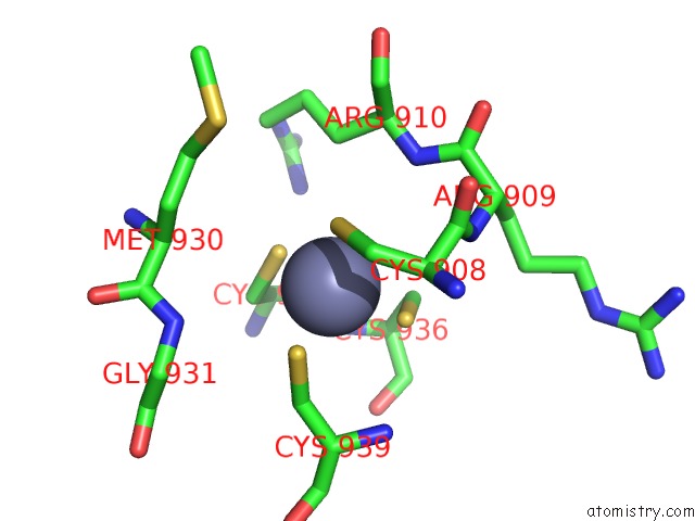
Mono view
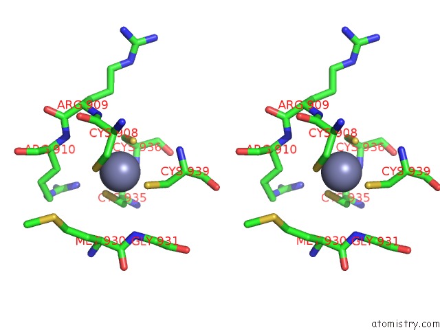
Stereo pair view
Mono view
Stereo pair view
A full contact list of Zinc with other atoms in the Zn binding
site number 1 of Crystal Structure of Werner Syndrome Helicase within 5.0Å range:
|
Zinc binding site 2 out of 2 in 6yhr
Go back to
Zinc binding site 2 out
of 2 in the Crystal Structure of Werner Syndrome Helicase
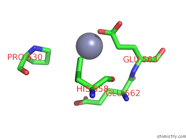
Mono view
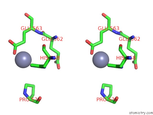
Stereo pair view
Mono view
Stereo pair view
A full contact list of Zinc with other atoms in the Zn binding
site number 2 of Crystal Structure of Werner Syndrome Helicase within 5.0Å range:
|
Reference:
J.A.Newman,
A.E.Gavard,
P.Savitsky,
F.Von Delft,
C.H.Arrowsmith,
A.Edwards,
C.Bountra,
O.Gileadi.
Crystal Structure of Werner Syndrome Helicase To Be Published.
Page generated: Thu Aug 21 21:28:27 2025
Last articles
Zn in 7K9AZn in 7K95
Zn in 7K99
Zn in 7K84
Zn in 7K7G
Zn in 7K7T
Zn in 7K6Z
Zn in 7K6X
Zn in 7K6U
Zn in 7K6T