Zinc »
PDB 6wq3-6x4y »
6wuq »
Zinc in PDB 6wuq: Crystal Structure of AJIA1 in Apo Form
Protein crystallography data
The structure of Crystal Structure of AJIA1 in Apo Form, PDB code: 6wuq
was solved by
F.C.R.Paiva,
K.Chan,
P.Leadlay,
M.V.B.Dias,
with X-Ray Crystallography technique. A brief refinement statistics is given in the table below:
| Resolution Low / High (Å) | 33.82 / 2.00 |
| Space group | P 31 2 1 |
| Cell size a, b, c (Å), α, β, γ (°) | 128.777, 128.777, 101.449, 90.00, 90.00, 120.00 |
| R / Rfree (%) | 18.8 / 22.4 |
Other elements in 6wuq:
The structure of Crystal Structure of AJIA1 in Apo Form also contains other interesting chemical elements:
| Magnesium | (Mg) | 2 atoms |
Zinc Binding Sites:
The binding sites of Zinc atom in the Crystal Structure of AJIA1 in Apo Form
(pdb code 6wuq). This binding sites where shown within
5.0 Angstroms radius around Zinc atom.
In total 2 binding sites of Zinc where determined in the Crystal Structure of AJIA1 in Apo Form, PDB code: 6wuq:
Jump to Zinc binding site number: 1; 2;
In total 2 binding sites of Zinc where determined in the Crystal Structure of AJIA1 in Apo Form, PDB code: 6wuq:
Jump to Zinc binding site number: 1; 2;
Zinc binding site 1 out of 2 in 6wuq
Go back to
Zinc binding site 1 out
of 2 in the Crystal Structure of AJIA1 in Apo Form
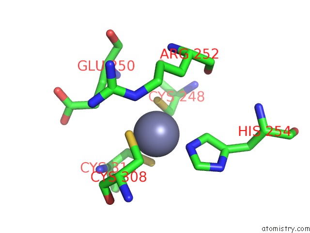
Mono view
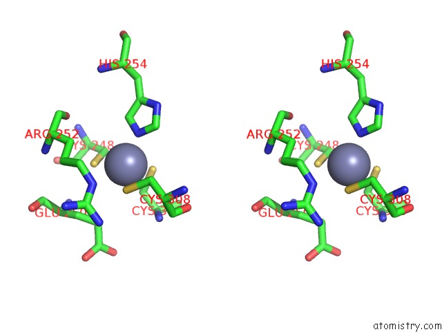
Stereo pair view
Mono view
Stereo pair view
A full contact list of Zinc with other atoms in the Zn binding
site number 1 of Crystal Structure of AJIA1 in Apo Form within 5.0Å range:
|
Zinc binding site 2 out of 2 in 6wuq
Go back to
Zinc binding site 2 out
of 2 in the Crystal Structure of AJIA1 in Apo Form
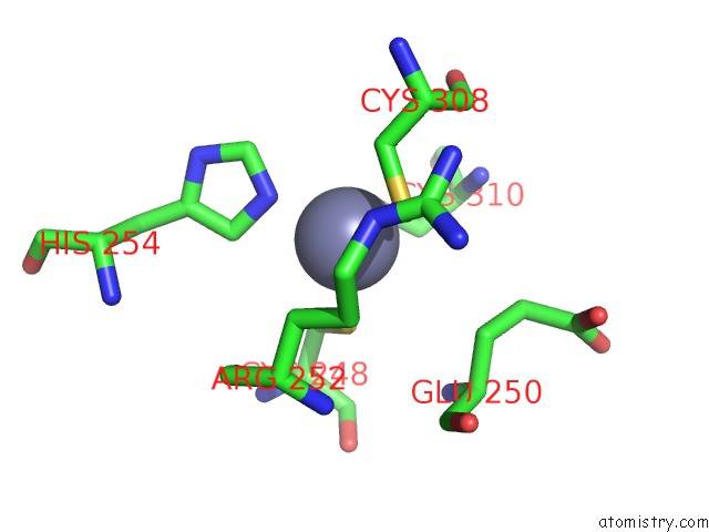
Mono view
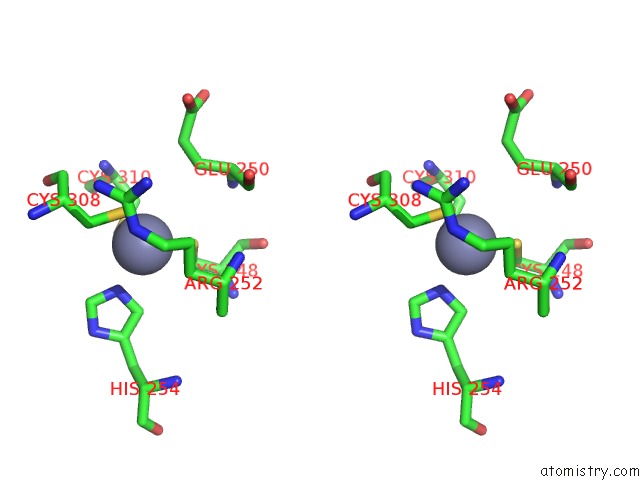
Stereo pair view
Mono view
Stereo pair view
A full contact list of Zinc with other atoms in the Zn binding
site number 2 of Crystal Structure of AJIA1 in Apo Form within 5.0Å range:
|
Reference:
F.C.R.Paiva,
K.Chan,
M.Samborskyy,
A.M.Silber,
P.Leadlay,
M.V.B.Dias.
The Crystal Structure of AJIA1 Reveals A Novel Structural Motion Mechanism in the Adenylate-Forming Enzyme Family Acta Crystallogr.,Sect.D V. 76 1201 2020.
ISSN: ESSN 1399-0047
DOI: 10.1107/S2059798320013431
Page generated: Tue Oct 29 10:24:09 2024
ISSN: ESSN 1399-0047
DOI: 10.1107/S2059798320013431
Last articles
Zn in 9MJ5Zn in 9HNW
Zn in 9G0L
Zn in 9FNE
Zn in 9DZN
Zn in 9E0I
Zn in 9D32
Zn in 9DAK
Zn in 8ZXC
Zn in 8ZUF