Zinc »
PDB 6v77-6vpc »
6vji »
Zinc in PDB 6vji: Structure of Mammalian NEIL2 From Monodelphis Domestica
Protein crystallography data
The structure of Structure of Mammalian NEIL2 From Monodelphis Domestica, PDB code: 6vji
was solved by
B.E.Eckenroth,
S.Doublie,
with X-Ray Crystallography technique. A brief refinement statistics is given in the table below:
| Resolution Low / High (Å) | 37.96 / 2.54 |
| Space group | P 32 |
| Cell size a, b, c (Å), α, β, γ (°) | 67.887, 67.887, 149.130, 90.00, 90.00, 120.00 |
| R / Rfree (%) | 25 / 27.5 |
Zinc Binding Sites:
The binding sites of Zinc atom in the Structure of Mammalian NEIL2 From Monodelphis Domestica
(pdb code 6vji). This binding sites where shown within
5.0 Angstroms radius around Zinc atom.
In total 2 binding sites of Zinc where determined in the Structure of Mammalian NEIL2 From Monodelphis Domestica, PDB code: 6vji:
Jump to Zinc binding site number: 1; 2;
In total 2 binding sites of Zinc where determined in the Structure of Mammalian NEIL2 From Monodelphis Domestica, PDB code: 6vji:
Jump to Zinc binding site number: 1; 2;
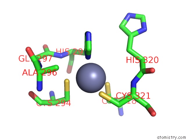
Zinc binding site 1 out of 2 in 6vji
Go back to
Zinc binding site 1 out
of 2 in the Structure of Mammalian NEIL2 From Monodelphis Domestica

Mono view

Stereo pair view
Mono view
Stereo pair view
A full contact list of Zinc with other atoms in the Zn binding
site number 1 of Structure of Mammalian NEIL2 From Monodelphis Domestica within 5.0Å range:
|
Zinc binding site 2 out of 2 in 6vji
Go back to
Zinc binding site 2 out
of 2 in the Structure of Mammalian NEIL2 From Monodelphis Domestica
Mono view

Stereo pair view
Mono view
Stereo pair view
A full contact list of Zinc with other atoms in the Zn binding
site number 2 of Structure of Mammalian NEIL2 From Monodelphis Domestica within 5.0Å range:
|
Reference:
B.E.Eckenroth,
V.B.Cao,
A.M.Averill,
J.A.Dragon,
S.Doublie.
Unique Structural Features of Mammalian NEIL2 Dna Glycosylase Prime Its Activity For Diverse Dna Substrates and Environments. Structure 2020.
ISSN: ISSN 0969-2126
PubMed: 32846144
DOI: 10.1016/J.STR.2020.08.001
Page generated: Tue Oct 29 09:03:19 2024
ISSN: ISSN 0969-2126
PubMed: 32846144
DOI: 10.1016/J.STR.2020.08.001
Last articles
Zn in 9MJ5Zn in 9HNW
Zn in 9G0L
Zn in 9FNE
Zn in 9DZN
Zn in 9E0I
Zn in 9D32
Zn in 9DAK
Zn in 8ZXC
Zn in 8ZUF