Zinc »
PDB 6tyh-6uca »
6u3u »
Zinc in PDB 6u3u: Crystal Structure of Shiga Toxin 2K
Protein crystallography data
The structure of Crystal Structure of Shiga Toxin 2K, PDB code: 6u3u
was solved by
Y.Z.Zhang,
X.H.He,
with X-Ray Crystallography technique. A brief refinement statistics is given in the table below:
| Resolution Low / High (Å) | 52.08 / 2.29 |
| Space group | P 1 21 1 |
| Cell size a, b, c (Å), α, β, γ (°) | 57.178, 157.022, 107.407, 90.00, 94.61, 90.00 |
| R / Rfree (%) | 19.7 / 22.5 |
Zinc Binding Sites:
The binding sites of Zinc atom in the Crystal Structure of Shiga Toxin 2K
(pdb code 6u3u). This binding sites where shown within
5.0 Angstroms radius around Zinc atom.
In total only one binding site of Zinc was determined in the Crystal Structure of Shiga Toxin 2K, PDB code: 6u3u:
In total only one binding site of Zinc was determined in the Crystal Structure of Shiga Toxin 2K, PDB code: 6u3u:
Zinc binding site 1 out of 1 in 6u3u
Go back to
Zinc binding site 1 out
of 1 in the Crystal Structure of Shiga Toxin 2K
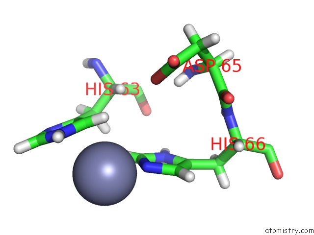
Mono view
Stereo pair view
Mono view
Stereo pair view
A full contact list of Zinc with other atoms in the Zn binding
site number 1 of Crystal Structure of Shiga Toxin 2K within 5.0Å range:
|
Reference:
A.C.Hughes,
Y.Zhang,
X.Bai,
Y.Xiong,
Y.Wang,
X.Yang,
Q.Xu,
X.He.
Structural and Functional Characterization of STX2K, A New Subtype of Shiga Toxin 2. Microorganisms V. 8 2019.
ISSN: ISSN 2076-2607
PubMed: 31861375
DOI: 10.3390/MICROORGANISMS8010004
Page generated: Tue Oct 29 08:23:38 2024
ISSN: ISSN 2076-2607
PubMed: 31861375
DOI: 10.3390/MICROORGANISMS8010004
Last articles
Zn in 9MJ5Zn in 9HNW
Zn in 9G0L
Zn in 9FNE
Zn in 9DZN
Zn in 9E0I
Zn in 9D32
Zn in 9DAK
Zn in 8ZXC
Zn in 8ZUF