Zinc »
PDB 6rwo-6s6b »
6rxp »
Zinc in PDB 6rxp: Crystal Structure of Cobb AC2 (A76G,I131C,V162A) in Complex with H4K16-Crotonyl Peptide
Protein crystallography data
The structure of Crystal Structure of Cobb AC2 (A76G,I131C,V162A) in Complex with H4K16-Crotonyl Peptide, PDB code: 6rxp
was solved by
M.Spinck,
R.Gasper,
H.Neumann,
with X-Ray Crystallography technique. A brief refinement statistics is given in the table below:
| Resolution Low / High (Å) | 47.95 / 1.80 |
| Space group | P 21 21 21 |
| Cell size a, b, c (Å), α, β, γ (°) | 57.560, 91.890, 95.900, 90.00, 90.00, 90.00 |
| R / Rfree (%) | 19.2 / 23.4 |
Zinc Binding Sites:
The binding sites of Zinc atom in the Crystal Structure of Cobb AC2 (A76G,I131C,V162A) in Complex with H4K16-Crotonyl Peptide
(pdb code 6rxp). This binding sites where shown within
5.0 Angstroms radius around Zinc atom.
In total 2 binding sites of Zinc where determined in the Crystal Structure of Cobb AC2 (A76G,I131C,V162A) in Complex with H4K16-Crotonyl Peptide, PDB code: 6rxp:
Jump to Zinc binding site number: 1; 2;
In total 2 binding sites of Zinc where determined in the Crystal Structure of Cobb AC2 (A76G,I131C,V162A) in Complex with H4K16-Crotonyl Peptide, PDB code: 6rxp:
Jump to Zinc binding site number: 1; 2;
Zinc binding site 1 out of 2 in 6rxp
Go back to
Zinc binding site 1 out
of 2 in the Crystal Structure of Cobb AC2 (A76G,I131C,V162A) in Complex with H4K16-Crotonyl Peptide
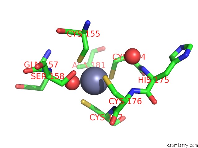
Mono view
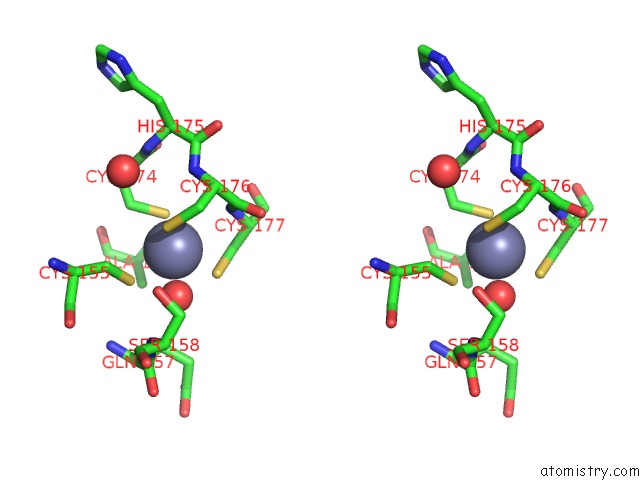
Stereo pair view
Mono view
Stereo pair view
A full contact list of Zinc with other atoms in the Zn binding
site number 1 of Crystal Structure of Cobb AC2 (A76G,I131C,V162A) in Complex with H4K16-Crotonyl Peptide within 5.0Å range:
|
Zinc binding site 2 out of 2 in 6rxp
Go back to
Zinc binding site 2 out
of 2 in the Crystal Structure of Cobb AC2 (A76G,I131C,V162A) in Complex with H4K16-Crotonyl Peptide
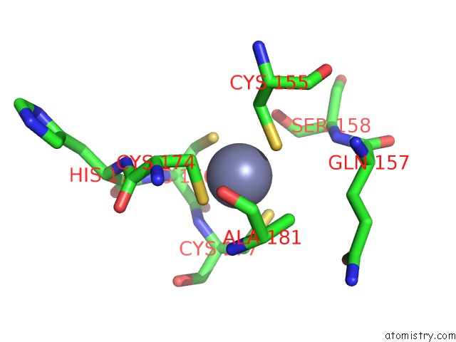
Mono view
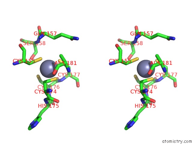
Stereo pair view
Mono view
Stereo pair view
A full contact list of Zinc with other atoms in the Zn binding
site number 2 of Crystal Structure of Cobb AC2 (A76G,I131C,V162A) in Complex with H4K16-Crotonyl Peptide within 5.0Å range:
|
Reference:
M.Spinck,
P.Neumann-Staubitz,
M.Ecke,
R.Gasper,
H.Neumann.
Evolved, Selective Erasers of Distinct Lysine Acylations. Angew.Chem.Int.Ed.Engl. 2020.
ISSN: ESSN 1521-3773
PubMed: 32187803
DOI: 10.1002/ANIE.202002899
Page generated: Tue Oct 29 06:54:08 2024
ISSN: ESSN 1521-3773
PubMed: 32187803
DOI: 10.1002/ANIE.202002899
Last articles
Zn in 9MJ5Zn in 9HNW
Zn in 9G0L
Zn in 9FNE
Zn in 9DZN
Zn in 9E0I
Zn in 9D32
Zn in 9DAK
Zn in 8ZXC
Zn in 8ZUF