Zinc »
PDB 6rwo-6s6b »
6rxd »
Zinc in PDB 6rxd: Crystal Structure of Bifidobacterium Longum Multiple Inositol Polyphosphate Phosphatase Apo Form
Protein crystallography data
The structure of Crystal Structure of Bifidobacterium Longum Multiple Inositol Polyphosphate Phosphatase Apo Form, PDB code: 6rxd
was solved by
A.W.H.Li,
C.A.Brearley,
A.M.Hemmings,
with X-Ray Crystallography technique. A brief refinement statistics is given in the table below:
| Resolution Low / High (Å) | 46.53 / 1.65 |
| Space group | P 1 |
| Cell size a, b, c (Å), α, β, γ (°) | 54.660, 72.940, 88.160, 71.61, 72.10, 77.17 |
| R / Rfree (%) | 15.3 / 17.7 |
Zinc Binding Sites:
The binding sites of Zinc atom in the Crystal Structure of Bifidobacterium Longum Multiple Inositol Polyphosphate Phosphatase Apo Form
(pdb code 6rxd). This binding sites where shown within
5.0 Angstroms radius around Zinc atom.
In total 4 binding sites of Zinc where determined in the Crystal Structure of Bifidobacterium Longum Multiple Inositol Polyphosphate Phosphatase Apo Form, PDB code: 6rxd:
Jump to Zinc binding site number: 1; 2; 3; 4;
In total 4 binding sites of Zinc where determined in the Crystal Structure of Bifidobacterium Longum Multiple Inositol Polyphosphate Phosphatase Apo Form, PDB code: 6rxd:
Jump to Zinc binding site number: 1; 2; 3; 4;
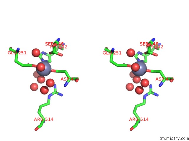
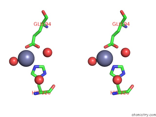
Zinc binding site 1 out of 4 in 6rxd
Go back to
Zinc binding site 1 out
of 4 in the Crystal Structure of Bifidobacterium Longum Multiple Inositol Polyphosphate Phosphatase Apo Form
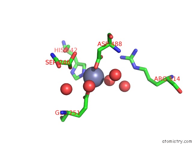
Mono view
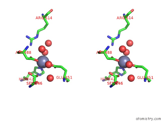
Stereo pair view
Mono view
Stereo pair view
A full contact list of Zinc with other atoms in the Zn binding
site number 1 of Crystal Structure of Bifidobacterium Longum Multiple Inositol Polyphosphate Phosphatase Apo Form within 5.0Å range:
|
Zinc binding site 2 out of 4 in 6rxd
Go back to
Zinc binding site 2 out
of 4 in the Crystal Structure of Bifidobacterium Longum Multiple Inositol Polyphosphate Phosphatase Apo Form

Mono view

Stereo pair view
Mono view
Stereo pair view
A full contact list of Zinc with other atoms in the Zn binding
site number 2 of Crystal Structure of Bifidobacterium Longum Multiple Inositol Polyphosphate Phosphatase Apo Form within 5.0Å range:
|
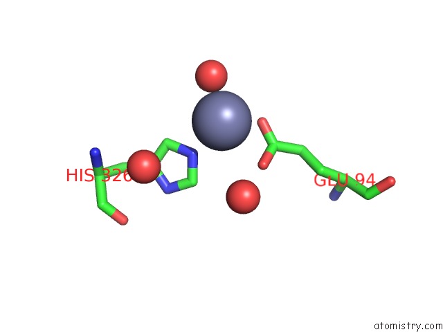
Zinc binding site 3 out of 4 in 6rxd
Go back to
Zinc binding site 3 out
of 4 in the Crystal Structure of Bifidobacterium Longum Multiple Inositol Polyphosphate Phosphatase Apo Form

Mono view
Stereo pair view
Mono view
Stereo pair view
A full contact list of Zinc with other atoms in the Zn binding
site number 3 of Crystal Structure of Bifidobacterium Longum Multiple Inositol Polyphosphate Phosphatase Apo Form within 5.0Å range:
|
Zinc binding site 4 out of 4 in 6rxd
Go back to
Zinc binding site 4 out
of 4 in the Crystal Structure of Bifidobacterium Longum Multiple Inositol Polyphosphate Phosphatase Apo Form
Mono view
Stereo pair view
Mono view
Stereo pair view
A full contact list of Zinc with other atoms in the Zn binding
site number 4 of Crystal Structure of Bifidobacterium Longum Multiple Inositol Polyphosphate Phosphatase Apo Form within 5.0Å range:
|
Reference:
I.M.Acquistapace,
A.W.H.Li,
M.A.Z.Zietek,
C.A.Brearley,
A.M.Hemmings.
Crystal Structures of Bifidobacterium Longum Multiple Inositol Phosphate Phosphatase Reveal An Alpha-Domain Induced-Fit Mechanism To Be Published.
Page generated: Tue Oct 29 06:49:54 2024
Last articles
Zn in 9MJ5Zn in 9HNW
Zn in 9G0L
Zn in 9FNE
Zn in 9DZN
Zn in 9E0I
Zn in 9D32
Zn in 9DAK
Zn in 8ZXC
Zn in 8ZUF