Zinc »
PDB 6rpn-6rwn »
6rvf »
Zinc in PDB 6rvf: Crystal Structure of Hca II in Complex with Urea, N-(1,3-Dihydro-1- Hydroxy-2,1-Benzoxaborol-6-Yl)-N'-Phenyl
Enzymatic activity of Crystal Structure of Hca II in Complex with Urea, N-(1,3-Dihydro-1- Hydroxy-2,1-Benzoxaborol-6-Yl)-N'-Phenyl
All present enzymatic activity of Crystal Structure of Hca II in Complex with Urea, N-(1,3-Dihydro-1- Hydroxy-2,1-Benzoxaborol-6-Yl)-N'-Phenyl:
4.2.1.1;
4.2.1.1;
Protein crystallography data
The structure of Crystal Structure of Hca II in Complex with Urea, N-(1,3-Dihydro-1- Hydroxy-2,1-Benzoxaborol-6-Yl)-N'-Phenyl, PDB code: 6rvf
was solved by
A.Di Fiore,
G.De Simone,
with X-Ray Crystallography technique. A brief refinement statistics is given in the table below:
| Resolution Low / High (Å) | 24.60 / 2.07 |
| Space group | P 1 21 1 |
| Cell size a, b, c (Å), α, β, γ (°) | 42.380, 41.380, 71.940, 90.00, 104.18, 90.00 |
| R / Rfree (%) | 18.4 / 22.9 |
Zinc Binding Sites:
The binding sites of Zinc atom in the Crystal Structure of Hca II in Complex with Urea, N-(1,3-Dihydro-1- Hydroxy-2,1-Benzoxaborol-6-Yl)-N'-Phenyl
(pdb code 6rvf). This binding sites where shown within
5.0 Angstroms radius around Zinc atom.
In total only one binding site of Zinc was determined in the Crystal Structure of Hca II in Complex with Urea, N-(1,3-Dihydro-1- Hydroxy-2,1-Benzoxaborol-6-Yl)-N'-Phenyl, PDB code: 6rvf:
In total only one binding site of Zinc was determined in the Crystal Structure of Hca II in Complex with Urea, N-(1,3-Dihydro-1- Hydroxy-2,1-Benzoxaborol-6-Yl)-N'-Phenyl, PDB code: 6rvf:
Zinc binding site 1 out of 1 in 6rvf
Go back to
Zinc binding site 1 out
of 1 in the Crystal Structure of Hca II in Complex with Urea, N-(1,3-Dihydro-1- Hydroxy-2,1-Benzoxaborol-6-Yl)-N'-Phenyl
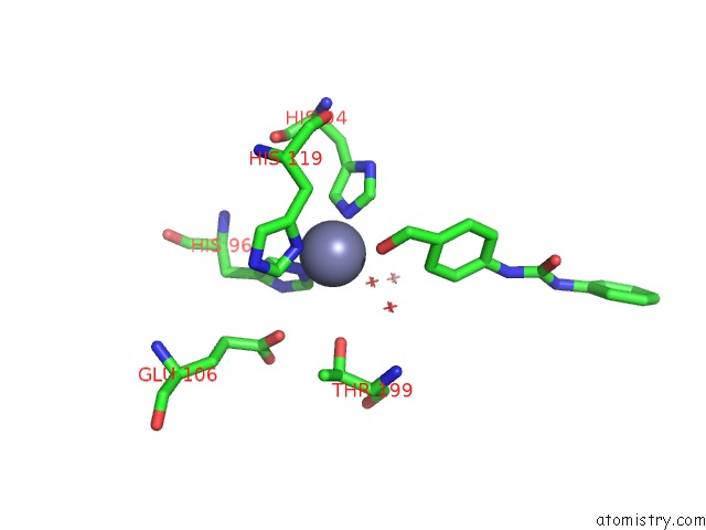
Mono view
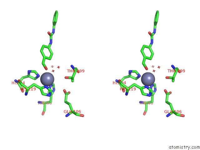
Stereo pair view
Mono view
Stereo pair view
A full contact list of Zinc with other atoms in the Zn binding
site number 1 of Crystal Structure of Hca II in Complex with Urea, N-(1,3-Dihydro-1- Hydroxy-2,1-Benzoxaborol-6-Yl)-N'-Phenyl within 5.0Å range:
|
Reference:
E.Langella,
V.Alterio,
K.D'ambrosio,
R.Cadoni,
J.Y.Winum,
C.T.Supuran,
S.M.Monti,
G.De Simone,
A.Di Fiore.
Exploring Benzoxaborole Derivatives As Carbonic Anhydrase Inhibitors: A Structural and Computational Analysis Reveals Their Conformational Variability As A Tool to Increase Enzyme Selectivity. J Enzyme Inhib Med Chem V. 34 1498 2019.
ISSN: ESSN 1475-6374
PubMed: 31423863
DOI: 10.1080/14756366.2019.1653291
Page generated: Tue Oct 29 06:47:53 2024
ISSN: ESSN 1475-6374
PubMed: 31423863
DOI: 10.1080/14756366.2019.1653291
Last articles
Zn in 9MJ5Zn in 9HNW
Zn in 9G0L
Zn in 9FNE
Zn in 9DZN
Zn in 9E0I
Zn in 9D32
Zn in 9DAK
Zn in 8ZXC
Zn in 8ZUF