Zinc »
PDB 6r06-6r54 »
6r4u »
Zinc in PDB 6r4u: Crystal Structure of the PRI1 Subunit of Human Primase Bound to Fludarabine Triphosphate
Protein crystallography data
The structure of Crystal Structure of the PRI1 Subunit of Human Primase Bound to Fludarabine Triphosphate, PDB code: 6r4u
was solved by
M.L.Kilkenny,
L.Pellegrini,
with X-Ray Crystallography technique. A brief refinement statistics is given in the table below:
| Resolution Low / High (Å) | 46.13 / 2.20 |
| Space group | C 2 2 21 |
| Cell size a, b, c (Å), α, β, γ (°) | 110.830, 117.570, 148.800, 90.00, 90.00, 90.00 |
| R / Rfree (%) | 19.6 / 22.3 |
Other elements in 6r4u:
The structure of Crystal Structure of the PRI1 Subunit of Human Primase Bound to Fludarabine Triphosphate also contains other interesting chemical elements:
| Fluorine | (F) | 2 atoms |
| Manganese | (Mn) | 4 atoms |
Zinc Binding Sites:
The binding sites of Zinc atom in the Crystal Structure of the PRI1 Subunit of Human Primase Bound to Fludarabine Triphosphate
(pdb code 6r4u). This binding sites where shown within
5.0 Angstroms radius around Zinc atom.
In total 2 binding sites of Zinc where determined in the Crystal Structure of the PRI1 Subunit of Human Primase Bound to Fludarabine Triphosphate, PDB code: 6r4u:
Jump to Zinc binding site number: 1; 2;
In total 2 binding sites of Zinc where determined in the Crystal Structure of the PRI1 Subunit of Human Primase Bound to Fludarabine Triphosphate, PDB code: 6r4u:
Jump to Zinc binding site number: 1; 2;
Zinc binding site 1 out of 2 in 6r4u
Go back to
Zinc binding site 1 out
of 2 in the Crystal Structure of the PRI1 Subunit of Human Primase Bound to Fludarabine Triphosphate
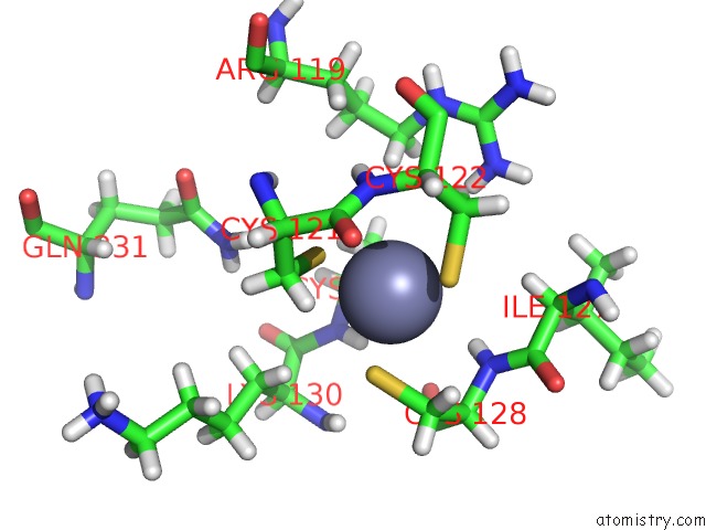
Mono view
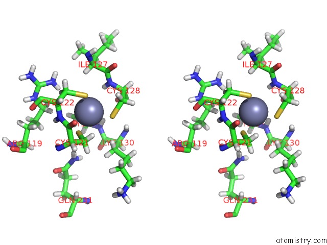
Stereo pair view
Mono view
Stereo pair view
A full contact list of Zinc with other atoms in the Zn binding
site number 1 of Crystal Structure of the PRI1 Subunit of Human Primase Bound to Fludarabine Triphosphate within 5.0Å range:
|
Zinc binding site 2 out of 2 in 6r4u
Go back to
Zinc binding site 2 out
of 2 in the Crystal Structure of the PRI1 Subunit of Human Primase Bound to Fludarabine Triphosphate
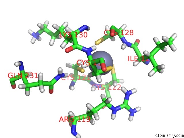
Mono view
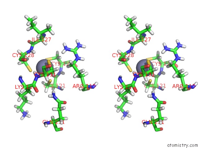
Stereo pair view
Mono view
Stereo pair view
A full contact list of Zinc with other atoms in the Zn binding
site number 2 of Crystal Structure of the PRI1 Subunit of Human Primase Bound to Fludarabine Triphosphate within 5.0Å range:
|
Reference:
S.Holzer,
N.J.Rzechorzek,
I.R.Short,
M.Jenkyn-Bedford,
L.Pellegrini,
M.L.Kilkenny.
Structural Basis For Inhibition of Human Primase By Arabinofuranosyl Nucleoside Analogues Fludarabine and Vidarabine. Acs Chem.Biol. V. 14 1904 2019.
ISSN: ESSN 1554-8937
PubMed: 31479243
DOI: 10.1021/ACSCHEMBIO.9B00367
Page generated: Tue Oct 29 05:58:05 2024
ISSN: ESSN 1554-8937
PubMed: 31479243
DOI: 10.1021/ACSCHEMBIO.9B00367
Last articles
Zn in 9MJ5Zn in 9HNW
Zn in 9G0L
Zn in 9FNE
Zn in 9DZN
Zn in 9E0I
Zn in 9D32
Zn in 9DAK
Zn in 8ZXC
Zn in 8ZUF