Zinc »
PDB 6qeb-6r05 »
6qqm »
Zinc in PDB 6qqm: Crystal Structure of the Alpha Carbonic Anhydrase From Schistosoma Mansoni
Enzymatic activity of Crystal Structure of the Alpha Carbonic Anhydrase From Schistosoma Mansoni
All present enzymatic activity of Crystal Structure of the Alpha Carbonic Anhydrase From Schistosoma Mansoni:
4.2.1.1;
4.2.1.1;
Protein crystallography data
The structure of Crystal Structure of the Alpha Carbonic Anhydrase From Schistosoma Mansoni, PDB code: 6qqm
was solved by
M.Ferraroni,
A.Angeli,
C.T.Supuran,
with X-Ray Crystallography technique. A brief refinement statistics is given in the table below:
| Resolution Low / High (Å) | 39.60 / 1.75 |
| Space group | P 32 2 1 |
| Cell size a, b, c (Å), α, β, γ (°) | 103.040, 103.040, 132.560, 90.00, 90.00, 120.00 |
| R / Rfree (%) | 17 / 20.4 |
Zinc Binding Sites:
The binding sites of Zinc atom in the Crystal Structure of the Alpha Carbonic Anhydrase From Schistosoma Mansoni
(pdb code 6qqm). This binding sites where shown within
5.0 Angstroms radius around Zinc atom.
In total 2 binding sites of Zinc where determined in the Crystal Structure of the Alpha Carbonic Anhydrase From Schistosoma Mansoni, PDB code: 6qqm:
Jump to Zinc binding site number: 1; 2;
In total 2 binding sites of Zinc where determined in the Crystal Structure of the Alpha Carbonic Anhydrase From Schistosoma Mansoni, PDB code: 6qqm:
Jump to Zinc binding site number: 1; 2;
Zinc binding site 1 out of 2 in 6qqm
Go back to
Zinc binding site 1 out
of 2 in the Crystal Structure of the Alpha Carbonic Anhydrase From Schistosoma Mansoni
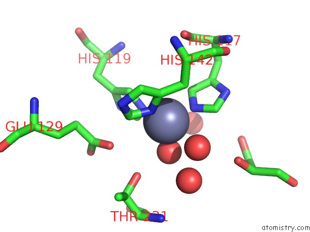
Mono view
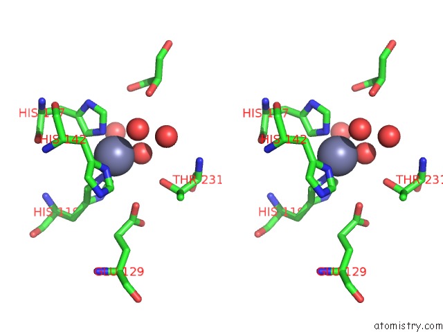
Stereo pair view
Mono view
Stereo pair view
A full contact list of Zinc with other atoms in the Zn binding
site number 1 of Crystal Structure of the Alpha Carbonic Anhydrase From Schistosoma Mansoni within 5.0Å range:
|
Zinc binding site 2 out of 2 in 6qqm
Go back to
Zinc binding site 2 out
of 2 in the Crystal Structure of the Alpha Carbonic Anhydrase From Schistosoma Mansoni
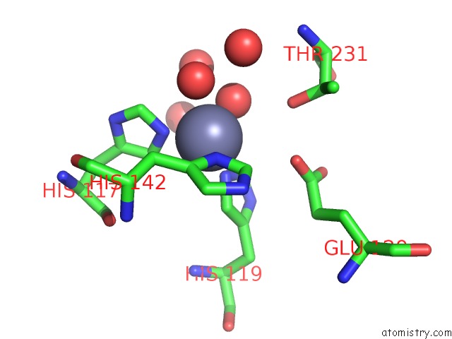
Mono view
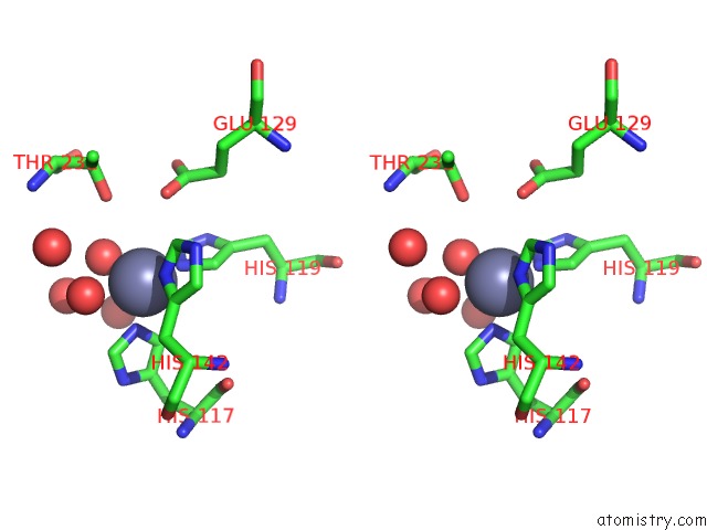
Stereo pair view
Mono view
Stereo pair view
A full contact list of Zinc with other atoms in the Zn binding
site number 2 of Crystal Structure of the Alpha Carbonic Anhydrase From Schistosoma Mansoni within 5.0Å range:
|
Reference:
A.A.Da'dara,
A.Angeli,
M.Ferraroni,
C.T.Supuran,
P.J.Skelly.
Crystal Structure and Chemical Inhibition of Essential Schistosome Host-Interactive Virulence Factor Carbonic Anhydrase Smca. Commun Biol V. 2 333 2019.
ISSN: ESSN 2399-3642
PubMed: 31508507
DOI: 10.1038/S42003-019-0578-0
Page generated: Tue Oct 29 05:44:25 2024
ISSN: ESSN 2399-3642
PubMed: 31508507
DOI: 10.1038/S42003-019-0578-0
Last articles
Zn in 9MJ5Zn in 9HNW
Zn in 9G0L
Zn in 9FNE
Zn in 9DZN
Zn in 9E0I
Zn in 9D32
Zn in 9DAK
Zn in 8ZXC
Zn in 8ZUF