Zinc »
PDB 6iu9-6j4e »
6j0d »
Zinc in PDB 6j0d: Crystal Structure of OSSUF4
Protein crystallography data
The structure of Crystal Structure of OSSUF4, PDB code: 6j0d
was solved by
B.Wang,
Q.Luo,
with X-Ray Crystallography technique. A brief refinement statistics is given in the table below:
| Resolution Low / High (Å) | 52.34 / 1.90 |
| Space group | P 41 21 2 |
| Cell size a, b, c (Å), α, β, γ (°) | 74.024, 74.024, 37.577, 90.00, 90.00, 90.00 |
| R / Rfree (%) | 19.6 / 23 |
Zinc Binding Sites:
The binding sites of Zinc atom in the Crystal Structure of OSSUF4
(pdb code 6j0d). This binding sites where shown within
5.0 Angstroms radius around Zinc atom.
In total 2 binding sites of Zinc where determined in the Crystal Structure of OSSUF4, PDB code: 6j0d:
Jump to Zinc binding site number: 1; 2;
In total 2 binding sites of Zinc where determined in the Crystal Structure of OSSUF4, PDB code: 6j0d:
Jump to Zinc binding site number: 1; 2;
Zinc binding site 1 out of 2 in 6j0d
Go back to
Zinc binding site 1 out
of 2 in the Crystal Structure of OSSUF4
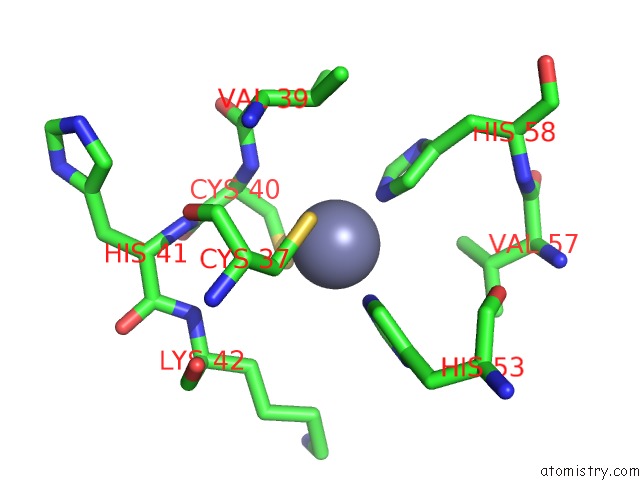
Mono view
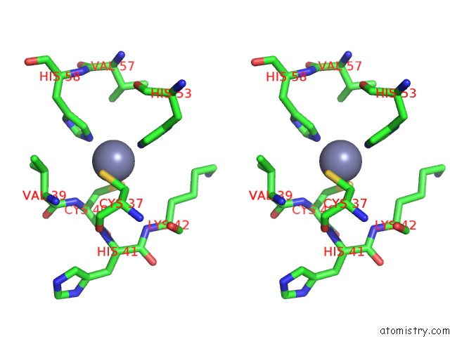
Stereo pair view
Mono view
Stereo pair view
A full contact list of Zinc with other atoms in the Zn binding
site number 1 of Crystal Structure of OSSUF4 within 5.0Å range:
|
Zinc binding site 2 out of 2 in 6j0d
Go back to
Zinc binding site 2 out
of 2 in the Crystal Structure of OSSUF4
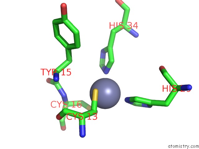
Mono view
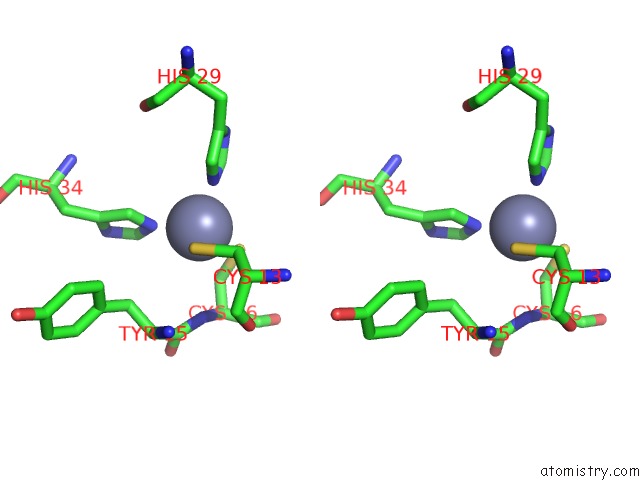
Stereo pair view
Mono view
Stereo pair view
A full contact list of Zinc with other atoms in the Zn binding
site number 2 of Crystal Structure of OSSUF4 within 5.0Å range:
|
Reference:
B.Liu,
Y.Liu,
B.Wang,
Q.Luo,
J.Shi,
J.Gan,
W.H.Shen,
Y.Yu,
A.Dong.
The Transcription Factor OSSUF4 Interacts with SDG725 in Promoting H3K36ME3 Establishment. Nat Commun V. 10 2999 2019.
ISSN: ESSN 2041-1723
PubMed: 31278262
DOI: 10.1038/S41467-019-10850-5
Page generated: Tue Oct 29 00:42:05 2024
ISSN: ESSN 2041-1723
PubMed: 31278262
DOI: 10.1038/S41467-019-10850-5
Last articles
Zn in 9MJ5Zn in 9HNW
Zn in 9G0L
Zn in 9FNE
Zn in 9DZN
Zn in 9E0I
Zn in 9D32
Zn in 9DAK
Zn in 8ZXC
Zn in 8ZUF