Atomistry » Zinc » PDB 6iu9-6j4e » 6ivk
Atomistry »
  Zinc »
    PDB 6iu9-6j4e »
      6ivk »

Zinc in PDB 6ivk: Crystal Structure of A Membrane Protein G175A

Protein crystallography data

The structure of Crystal Structure of A Membrane Protein G175A, PDB code: 6ivk was solved by A.Kittredge, F.Fukuda, Y.Zhang, T.Yang, with X-Ray Crystallography technique. A brief refinement statistics is given in the table below:

Resolution Low / High (Å) 48.64 / 2.65
Space group P 21 21 21
Cell size a, b, c (Å), α, β, γ (°) 113.548, 159.424, 161.476, 90.00, 90.00, 90.00
R / Rfree (%) 20.1 / 24.1

Other elements in 6ivk:

The structure of Crystal Structure of A Membrane Protein G175A also contains other interesting chemical elements:

Chlorine (Cl) 10 atoms

Zinc Binding Sites:

Pages:

>>> Page 1 <<< Page 2, Binding sites: 11 - 16;

Binding sites:

The binding sites of Zinc atom in the Crystal Structure of A Membrane Protein G175A (pdb code 6ivk). This binding sites where shown within 5.0 Angstroms radius around Zinc atom.
In total 16 binding sites of Zinc where determined in the Crystal Structure of A Membrane Protein G175A, PDB code: 6ivk:
Jump to Zinc binding site number: 1; 2; 3; 4; 5; 6; 7; 8; 9; 10;

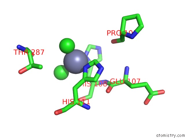
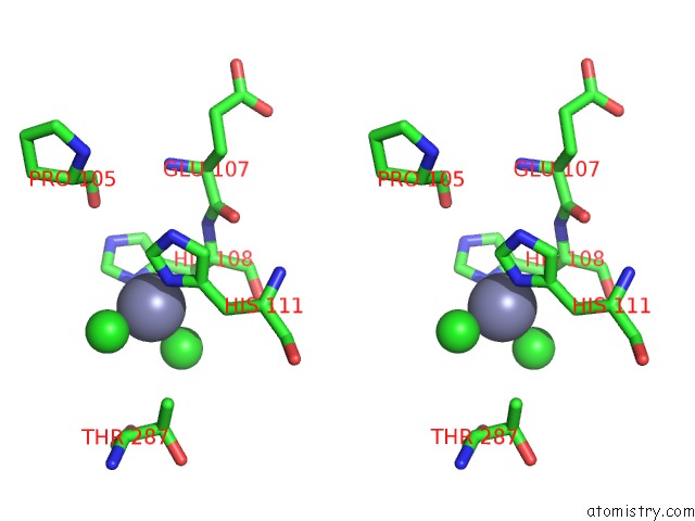
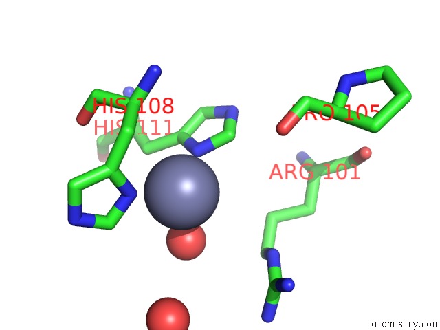
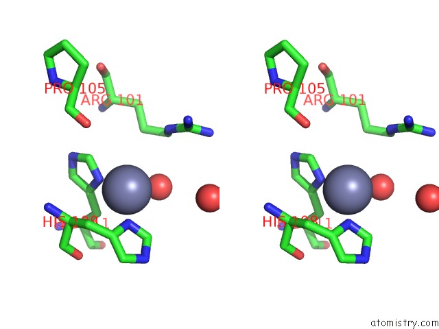
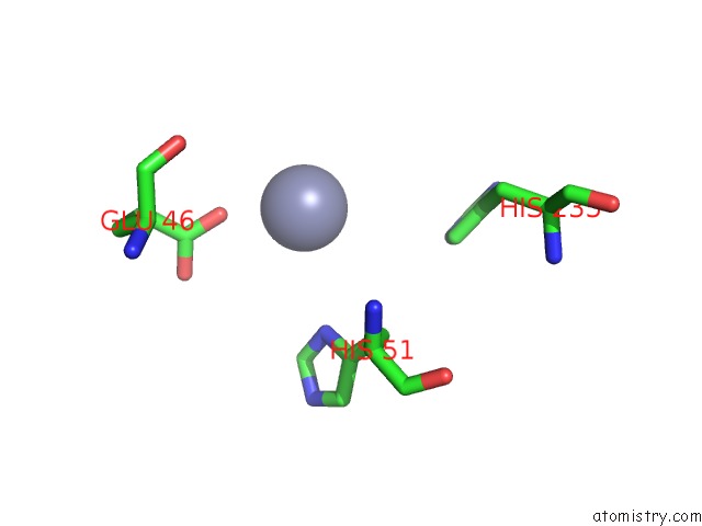
Zinc binding site 1 out of 16 in 6ivk

Go back to Zinc Binding Sites List in 6ivk
Zinc binding site 1 out of 16 in the Crystal Structure of A Membrane Protein G175A


Mono view


Stereo pair view

A full contact list of Zinc with other atoms in the Zn binding site number 1 of Crystal Structure of A Membrane Protein G175A within 5.0Å range:
probe atom residue distance (Å) B Occ
A:Zn301

b:58.9
occ:1.00
OE2 A:GLU191 1.9 71.6 1.0
NE2 E:HIS194 2.0 42.1 1.0
CL A:CL308 2.2 62.6 1.0
O A:HOH418 2.2 55.1 1.0
CD A:GLU191 2.7 62.0 1.0
OE1 A:GLU191 2.8 60.5 1.0
CE1 E:HIS194 3.0 42.7 1.0
CD2 E:HIS194 3.1 37.8 1.0
ND1 E:HIS194 4.1 46.0 1.0
CG A:GLU191 4.1 52.7 1.0
CG E:HIS194 4.2 41.3 1.0
CE A:LYS188 4.3 62.4 1.0
O E:HOH416 4.4 60.7 1.0
CD A:LYS188 4.4 55.5 1.0
CG2 A:ILE91 4.5 32.6 1.0
CB E:ASP190 4.7 50.6 1.0

Zinc binding site 2 out of 16 in 6ivk

Go back to Zinc Binding Sites List in 6ivk
Zinc binding site 2 out of 16 in the Crystal Structure of A Membrane Protein G175A


Mono view


Stereo pair view

A full contact list of Zinc with other atoms in the Zn binding site number 2 of Crystal Structure of A Membrane Protein G175A within 5.0Å range:
probe atom residue distance (Å) B Occ
A:Zn302

b:69.8
occ:1.00
CL B:CL304 2.0 81.2 1.0
NE2 A:HIS194 2.2 43.6 1.0
OE1 B:GLU191 2.3 66.5 1.0
O A:HOH423 2.4 53.4 1.0
OE2 B:GLU191 2.7 74.0 1.0
CD B:GLU191 2.8 69.2 1.0
CE1 A:HIS194 3.0 43.5 1.0
CD2 A:HIS194 3.2 37.5 1.0
CE B:LYS188 3.9 59.0 1.0
ND1 A:HIS194 4.2 43.1 1.0
CG B:GLU191 4.3 59.7 1.0
CG A:HIS194 4.3 39.4 1.0
OD2 A:ASP190 4.5 89.0 1.0
CG2 B:ILE91 4.5 32.3 1.0
CD B:LYS188 4.6 51.6 1.0
CB A:ASP190 4.7 51.6 1.0
NZ B:LYS188 4.8 74.5 1.0
CG A:ASP190 4.9 72.8 1.0

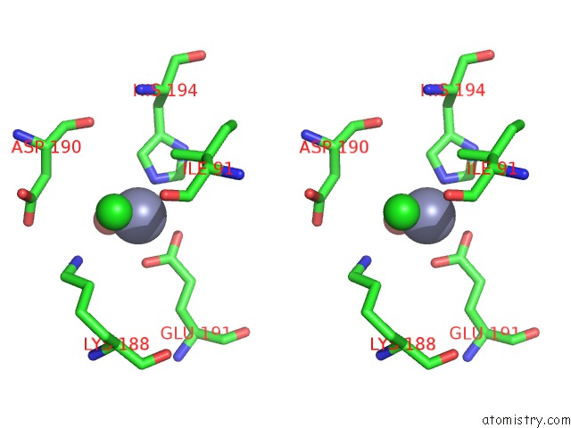
Zinc binding site 3 out of 16 in 6ivk

Go back to Zinc Binding Sites List in 6ivk
Zinc binding site 3 out of 16 in the Crystal Structure of A Membrane Protein G175A


Mono view


Stereo pair view

A full contact list of Zinc with other atoms in the Zn binding site number 3 of Crystal Structure of A Membrane Protein G175A within 5.0Å range:
probe atom residue distance (Å) B Occ
A:Zn303

b:63.9
occ:1.00
CL A:CL310 2.1 71.0 1.0
ND1 A:HIS111 2.1 49.2 1.0
ND1 A:HIS108 2.2 80.0 1.0
CL A:CL309 2.3 74.0 1.0
CE1 A:HIS111 3.0 53.6 1.0
CG A:HIS108 3.1 74.3 1.0
CE1 A:HIS108 3.1 77.4 1.0
CG A:HIS111 3.2 56.4 1.0
CB A:HIS108 3.3 66.0 1.0
CA A:HIS108 3.4 66.8 1.0
CB A:HIS111 3.5 56.5 1.0
NE2 A:HIS111 4.1 49.6 1.0
CD2 A:HIS108 4.2 70.5 1.0
NE2 A:HIS108 4.2 83.1 1.0
CD2 A:HIS111 4.2 54.1 1.0
N A:HIS108 4.3 67.8 1.0
O A:HIS108 4.5 68.7 1.0
C A:HIS108 4.5 67.3 1.0
CG2 A:THR287 4.5 59.8 1.0
O A:PRO105 4.8 63.8 1.0
C A:GLU107 4.8 66.3 1.0
CB A:THR287 4.9 74.8 1.0
O A:GLU107 4.9 72.1 1.0

Zinc binding site 4 out of 16 in 6ivk

Go back to Zinc Binding Sites List in 6ivk
Zinc binding site 4 out of 16 in the Crystal Structure of A Membrane Protein G175A


Mono view


Stereo pair view

A full contact list of Zinc with other atoms in the Zn binding site number 4 of Crystal Structure of A Membrane Protein G175A within 5.0Å range:
probe atom residue distance (Å) B Occ
A:Zn304

b:68.7
occ:1.00
OD2 A:ASP261 1.9 70.2 1.0
OD2 A:ASP269 2.1 57.3 1.0
OXT A:ACY305 2.6 88.7 1.0
CG A:ASP269 2.9 47.5 1.0
CG A:ASP261 3.0 71.2 1.0
OD1 A:ASP269 3.0 57.5 1.0
CB A:ASP261 3.5 60.4 1.0
C A:ACY305 3.5 100.0 1.0
CB A:ALA266 3.6 47.8 1.0
CA A:GLY264 3.8 39.4 1.0
O A:ASP261 3.8 50.5 1.0
OD1 A:ASP261 4.1 74.0 1.0
N A:ALA266 4.2 61.4 1.0
N A:THR265 4.3 54.0 1.0
CB A:ASP269 4.3 53.0 1.0
O A:ACY305 4.3 0.1 1.0
CH3 A:ACY305 4.3 86.7 1.0
C A:GLY264 4.3 48.8 1.0
CA A:ALA266 4.5 54.7 1.0
N A:GLY264 4.5 40.3 1.0
CA A:ASP261 4.6 60.6 1.0
C A:ASP261 4.6 55.6 1.0

Zinc binding site 5 out of 16 in 6ivk

Go back to Zinc Binding Sites List in 6ivk
Zinc binding site 5 out of 16 in the Crystal Structure of A Membrane Protein G175A


Mono view


Stereo pair view

A full contact list of Zinc with other atoms in the Zn binding site number 5 of Crystal Structure of A Membrane Protein G175A within 5.0Å range:
probe atom residue distance (Å) B Occ
B:Zn301

b:0.3
occ:1.00
O B:HOH427 2.2 72.5 1.0
ND1 B:HIS235 2.2 0.4 1.0
O B:HOH419 3.0 57.5 1.0
CG B:HIS235 3.1 0.2 1.0
CB B:HIS235 3.2 0.2 1.0
CE1 B:HIS235 3.2 91.4 1.0
CA B:HIS235 3.7 0.9 1.0
CD2 B:TYR42 3.9 87.7 1.0
CD2 B:HIS235 4.2 98.0 1.0
NE2 B:HIS235 4.3 88.3 1.0
CE2 B:TYR42 4.3 88.3 1.0
C B:HIS235 4.5 0.1 1.0
N B:HIS235 4.9 0.5 1.0
CG2 B:VAL231 4.9 69.7 1.0
N B:TYR236 4.9 0.7 1.0
CG B:TYR42 5.0 83.5 1.0

Zinc binding site 6 out of 16 in 6ivk

Go back to Zinc Binding Sites List in 6ivk
Zinc binding site 6 out of 16 in the Crystal Structure of A Membrane Protein G175A


Mono view


Stereo pair view

A full contact list of Zinc with other atoms in the Zn binding site number 6 of Crystal Structure of A Membrane Protein G175A within 5.0Å range:
probe atom residue distance (Å) B Occ
B:Zn302

b:55.4
occ:1.00
NE2 B:HIS290 1.9 56.0 1.0
ND1 B:HIS111 2.0 50.4 1.0
ND1 B:HIS108 2.2 61.4 1.0
OXT B:ACY303 2.2 53.2 1.0
CE1 B:HIS290 2.8 56.8 1.0
CE1 B:HIS111 2.8 50.5 1.0
C B:ACY303 3.0 65.3 1.0
O B:ACY303 3.0 57.7 1.0
CE1 B:HIS108 3.0 59.9 1.0
CD2 B:HIS290 3.0 54.6 1.0
CG B:HIS111 3.0 48.5 1.0
CG B:HIS108 3.2 55.4 1.0
CB B:HIS111 3.5 48.1 1.0
CB B:HIS108 3.6 48.2 1.0
CA B:HIS108 3.6 50.8 1.0
ND1 B:HIS290 4.0 52.7 1.0
NE2 B:HIS111 4.0 46.2 1.0
CG B:HIS290 4.1 59.1 1.0
CD2 B:HIS111 4.1 50.1 1.0
NE2 B:HIS108 4.2 54.5 1.0
N B:HIS108 4.2 49.5 1.0
CD2 B:HIS108 4.3 53.1 1.0
O B:PRO105 4.4 64.3 1.0
O B:GLY288 4.5 52.8 1.0
CH3 B:ACY303 4.5 52.3 1.0
CG B:ARG101 4.7 41.1 1.0
C B:HIS108 4.7 52.5 1.0
NE B:ARG101 4.8 57.7 1.0
C B:GLU107 4.8 52.5 1.0
O B:HIS108 4.8 54.9 1.0
O B:GLU107 4.9 57.1 1.0
CA B:HIS111 5.0 50.1 1.0

Zinc binding site 7 out of 16 in 6ivk

Go back to Zinc Binding Sites List in 6ivk
Zinc binding site 7 out of 16 in the Crystal Structure of A Membrane Protein G175A


Mono view


Stereo pair view

A full contact list of Zinc with other atoms in the Zn binding site number 7 of Crystal Structure of A Membrane Protein G175A within 5.0Å range:
probe atom residue distance (Å) B Occ
C:Zn301

b:55.7
occ:1.00
O C:HOH411 2.0 50.3 1.0
NE2 D:HIS194 2.0 42.1 1.0
CL C:CL306 2.2 57.6 1.0
OE1 C:GLU191 2.3 62.6 1.0
OE2 C:GLU191 2.4 64.4 1.0
CD C:GLU191 2.7 62.2 1.0
CE1 D:HIS194 3.0 44.5 1.0
CD2 D:HIS194 3.0 45.5 1.0
NZ C:LYS188 3.7 71.0 1.0
ND1 D:HIS194 4.1 46.6 1.0
CG D:HIS194 4.1 44.5 1.0
CG C:GLU191 4.2 54.3 1.0
CG2 C:ILE91 4.5 33.7 1.0
CE C:LYS188 4.9 61.3 1.0
CB D:ASP190 4.9 55.1 1.0
CD C:LYS188 4.9 54.4 1.0

Zinc binding site 8 out of 16 in 6ivk

Go back to Zinc Binding Sites List in 6ivk
Zinc binding site 8 out of 16 in the Crystal Structure of A Membrane Protein G175A


Mono view


Stereo pair view

A full contact list of Zinc with other atoms in the Zn binding site number 8 of Crystal Structure of A Membrane Protein G175A within 5.0Å range:
probe atom residue distance (Å) B Occ
C:Zn302

b:74.2
occ:1.00
CL C:CL305 2.0 84.8 1.0
OE1 E:GLU191 2.0 63.4 1.0
NE2 C:HIS194 2.0 43.7 1.0
O C:HOH415 2.5 58.8 1.0
OD1 C:ASP190 2.8 86.7 1.0
CD E:GLU191 2.9 61.8 1.0
CE1 C:HIS194 3.0 41.4 1.0
CD2 C:HIS194 3.0 39.7 1.0
OE2 E:GLU191 3.3 73.6 1.0
O E:HOH412 3.5 73.3 1.0
CG C:ASP190 3.7 71.7 1.0
OD2 C:ASP190 3.9 82.4 1.0
CE E:LYS188 4.1 60.6 1.0
ND1 C:HIS194 4.1 42.1 1.0
CG C:HIS194 4.2 39.6 1.0
CG E:GLU191 4.2 47.1 1.0
CG2 E:ILE91 4.7 33.3 1.0
CD E:LYS188 4.7 49.8 1.0

Zinc binding site 9 out of 16 in 6ivk

Go back to Zinc Binding Sites List in 6ivk
Zinc binding site 9 out of 16 in the Crystal Structure of A Membrane Protein G175A


Mono view


Stereo pair view

A full contact list of Zinc with other atoms in the Zn binding site number 9 of Crystal Structure of A Membrane Protein G175A within 5.0Å range:
probe atom residue distance (Å) B Occ
C:Zn303

b:58.8
occ:1.00
ND1 C:HIS111 1.9 55.4 1.0
ND1 C:HIS108 2.0 50.1 1.0
O C:HOH418 2.2 40.0 1.0
CE1 C:HIS111 2.9 53.9 1.0
CE1 C:HIS108 2.9 48.4 1.0
CG C:HIS111 3.0 53.5 1.0
CG C:HIS108 3.1 49.1 1.0
CB C:HIS111 3.3 51.8 1.0
CB C:HIS108 3.5 52.7 1.0
CA C:HIS108 3.7 59.5 1.0
NE2 C:HIS111 4.0 54.9 1.0
CD2 C:HIS111 4.1 52.0 1.0
NE2 C:HIS108 4.1 49.0 1.0
CD2 C:HIS108 4.2 43.2 1.0
NE C:ARG101 4.3 77.3 1.0
O C:HIS108 4.6 53.9 1.0
N C:HIS108 4.6 62.4 1.0
C C:HIS108 4.7 57.6 1.0
O C:PRO105 4.7 62.7 1.0
O C:HOH420 4.8 84.1 1.0
CG C:ARG101 4.8 57.4 1.0
CD C:ARG101 4.8 71.3 1.0
CA C:HIS111 4.9 51.4 1.0
CZ C:ARG101 4.9 71.5 1.0

Zinc binding site 10 out of 16 in 6ivk

Go back to Zinc Binding Sites List in 6ivk
Zinc binding site 10 out of 16 in the Crystal Structure of A Membrane Protein G175A


Mono view


Stereo pair view

A full contact list of Zinc with other atoms in the Zn binding site number 10 of Crystal Structure of A Membrane Protein G175A within 5.0Å range:
probe atom residue distance (Å) B Occ
C:Zn304

b:0.8
occ:1.00
OE2 C:GLU46 2.8 0.7 1.0
CD C:GLU46 3.9 0.2 1.0
ND1 C:HIS51 4.1 0.2 1.0
OE1 C:GLU46 4.3 0.9 1.0
CE1 C:HIS51 4.8 0.2 1.0
NE2 E:HIS235 4.9 0.8 1.0
CD2 E:HIS235 4.9 0.8 1.0

Reference:

C.Ji, A.Kittredge, A.Hopiavuori, N.Ward, S.Chen, Y.Fukuda, Y.Zhang, T.Yang. Dual CA2+-Dependent Gates in Human BESTROPHIN1 Underlie Disease-Causing Mechanisms of Gain-of-Function Mutations. Commun Biol V. 2 240 2019.
ISSN: ESSN 2399-3642
PubMed: 31263784
DOI: 10.1038/S42003-019-0433-3
Page generated: Tue Oct 29 00:20:49 2024

Last articles

Ar in 6QAR
Ar in 6R1Q
Ar in 7Q09
Ar in 7PWN
Ar in 7PUF
Ar in 6Q8R
Ar in 6ZMC
Ar in 6RA8
Ar in 6Q8S
Ar in 6IA3
© Copyright 2008-2020 by atomistry.com
Home   |    Site Map   |    Copyright   |    Contact us   |    Privacy