Zinc »
PDB 6i0w-6iiw »
6i6z »
Zinc in PDB 6i6z: Crystal Structure of the Human Carboxypeptidase A1 in Complex with the Phosphinic Inhibitor Acetyl-Tyr-Ala-Y(PO2CH2)-Homophe-Oh
Enzymatic activity of Crystal Structure of the Human Carboxypeptidase A1 in Complex with the Phosphinic Inhibitor Acetyl-Tyr-Ala-Y(PO2CH2)-Homophe-Oh
All present enzymatic activity of Crystal Structure of the Human Carboxypeptidase A1 in Complex with the Phosphinic Inhibitor Acetyl-Tyr-Ala-Y(PO2CH2)-Homophe-Oh:
3.4.17.1;
3.4.17.1;
Protein crystallography data
The structure of Crystal Structure of the Human Carboxypeptidase A1 in Complex with the Phosphinic Inhibitor Acetyl-Tyr-Ala-Y(PO2CH2)-Homophe-Oh, PDB code: 6i6z
was solved by
P.Gallego,
D.Reverter,
with X-Ray Crystallography technique. A brief refinement statistics is given in the table below:
| Resolution Low / High (Å) | 40.44 / 1.72 |
| Space group | P 21 21 21 |
| Cell size a, b, c (Å), α, β, γ (°) | 46.121, 84.109, 158.195, 90.00, 90.00, 90.00 |
| R / Rfree (%) | 15.8 / 18.3 |
Zinc Binding Sites:
The binding sites of Zinc atom in the Crystal Structure of the Human Carboxypeptidase A1 in Complex with the Phosphinic Inhibitor Acetyl-Tyr-Ala-Y(PO2CH2)-Homophe-Oh
(pdb code 6i6z). This binding sites where shown within
5.0 Angstroms radius around Zinc atom.
In total 2 binding sites of Zinc where determined in the Crystal Structure of the Human Carboxypeptidase A1 in Complex with the Phosphinic Inhibitor Acetyl-Tyr-Ala-Y(PO2CH2)-Homophe-Oh, PDB code: 6i6z:
Jump to Zinc binding site number: 1; 2;
In total 2 binding sites of Zinc where determined in the Crystal Structure of the Human Carboxypeptidase A1 in Complex with the Phosphinic Inhibitor Acetyl-Tyr-Ala-Y(PO2CH2)-Homophe-Oh, PDB code: 6i6z:
Jump to Zinc binding site number: 1; 2;
Zinc binding site 1 out of 2 in 6i6z
Go back to
Zinc binding site 1 out
of 2 in the Crystal Structure of the Human Carboxypeptidase A1 in Complex with the Phosphinic Inhibitor Acetyl-Tyr-Ala-Y(PO2CH2)-Homophe-Oh
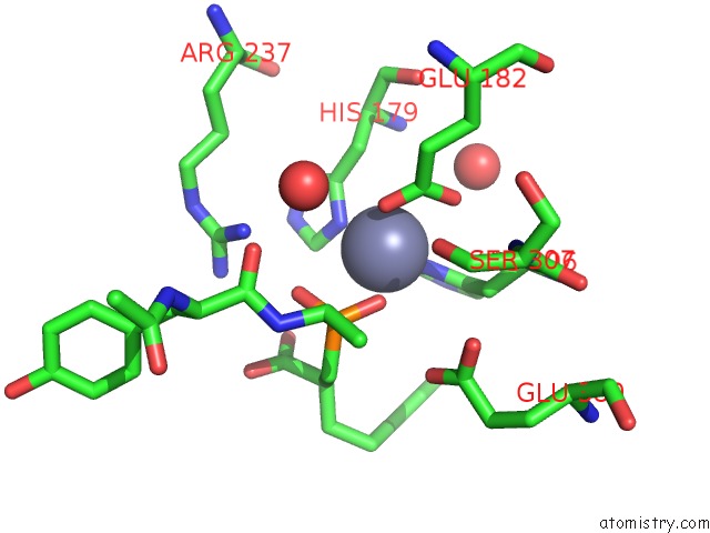
Mono view
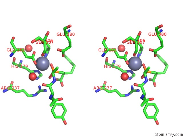
Stereo pair view
Mono view
Stereo pair view
A full contact list of Zinc with other atoms in the Zn binding
site number 1 of Crystal Structure of the Human Carboxypeptidase A1 in Complex with the Phosphinic Inhibitor Acetyl-Tyr-Ala-Y(PO2CH2)-Homophe-Oh within 5.0Å range:
|
Zinc binding site 2 out of 2 in 6i6z
Go back to
Zinc binding site 2 out
of 2 in the Crystal Structure of the Human Carboxypeptidase A1 in Complex with the Phosphinic Inhibitor Acetyl-Tyr-Ala-Y(PO2CH2)-Homophe-Oh
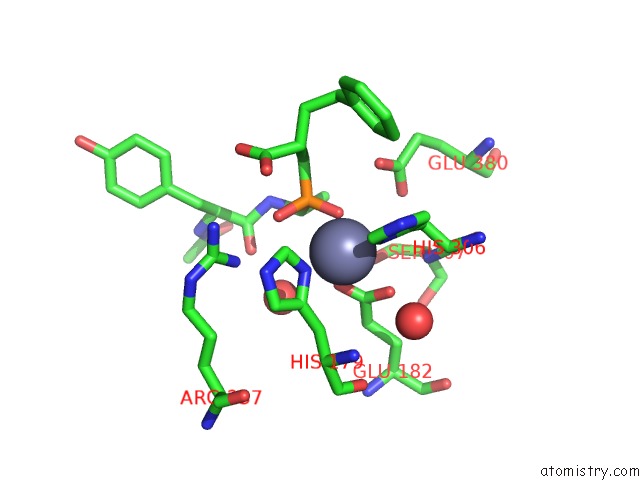
Mono view
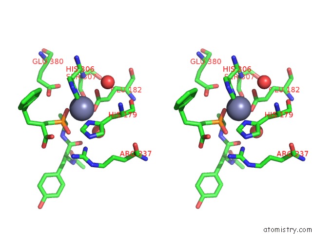
Stereo pair view
Mono view
Stereo pair view
A full contact list of Zinc with other atoms in the Zn binding
site number 2 of Crystal Structure of the Human Carboxypeptidase A1 in Complex with the Phosphinic Inhibitor Acetyl-Tyr-Ala-Y(PO2CH2)-Homophe-Oh within 5.0Å range:
|
Reference:
G.Covaleda,
P.Gallego,
J.Vendrell,
D.Georgiadis,
J.Lorenzo,
V.Dive,
F.X.Aviles,
D.Reverter,
L.Devel.
Synthesis and Structural/Functional Characterization of Selective M14 Metallocarboxypeptidase Inhibitors Based on Phosphinic Pseudopeptide Scaffold: Implications on the Design of Specific Optical Probes. J. Med. Chem. V. 62 1917 2019.
ISSN: ISSN 1520-4804
PubMed: 30688452
DOI: 10.1021/ACS.JMEDCHEM.8B01465
Page generated: Mon Oct 28 23:35:32 2024
ISSN: ISSN 1520-4804
PubMed: 30688452
DOI: 10.1021/ACS.JMEDCHEM.8B01465
Last articles
Zn in 9MJ5Zn in 9HNW
Zn in 9G0L
Zn in 9FNE
Zn in 9DZN
Zn in 9E0I
Zn in 9D32
Zn in 9DAK
Zn in 8ZXC
Zn in 8ZUF