Zinc »
PDB 6i0w-6iiw »
6i1d »
Zinc in PDB 6i1d: Structure of the YSH1-MPE1 Nuclease Complex From S.Cerevisiae
Protein crystallography data
The structure of Structure of the YSH1-MPE1 Nuclease Complex From S.Cerevisiae, PDB code: 6i1d
was solved by
C.H.Hill,
V.Boreikaite,
A.Kumar,
A.Casanal,
P.Kubik,
G.Degliesposti,
S.Maslen,
A.Mariani,
O.Von Loeffelholz,
M.Girbig,
M.Skehel,
L.A.Passmore,
with X-Ray Crystallography technique. A brief refinement statistics is given in the table below:
| Resolution Low / High (Å) | 62.14 / 2.28 |
| Space group | P 1 21 1 |
| Cell size a, b, c (Å), α, β, γ (°) | 43.380, 124.270, 63.450, 90.00, 103.21, 90.00 |
| R / Rfree (%) | 17.3 / 22.2 |
Zinc Binding Sites:
The binding sites of Zinc atom in the Structure of the YSH1-MPE1 Nuclease Complex From S.Cerevisiae
(pdb code 6i1d). This binding sites where shown within
5.0 Angstroms radius around Zinc atom.
In total 2 binding sites of Zinc where determined in the Structure of the YSH1-MPE1 Nuclease Complex From S.Cerevisiae, PDB code: 6i1d:
Jump to Zinc binding site number: 1; 2;
In total 2 binding sites of Zinc where determined in the Structure of the YSH1-MPE1 Nuclease Complex From S.Cerevisiae, PDB code: 6i1d:
Jump to Zinc binding site number: 1; 2;
Zinc binding site 1 out of 2 in 6i1d
Go back to
Zinc binding site 1 out
of 2 in the Structure of the YSH1-MPE1 Nuclease Complex From S.Cerevisiae
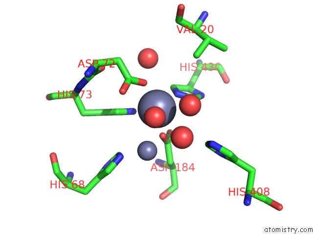
Mono view
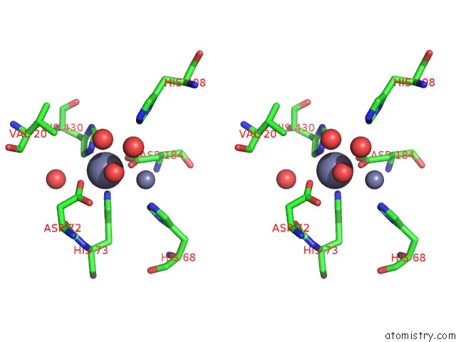
Stereo pair view
Mono view
Stereo pair view
A full contact list of Zinc with other atoms in the Zn binding
site number 1 of Structure of the YSH1-MPE1 Nuclease Complex From S.Cerevisiae within 5.0Å range:
|
Zinc binding site 2 out of 2 in 6i1d
Go back to
Zinc binding site 2 out
of 2 in the Structure of the YSH1-MPE1 Nuclease Complex From S.Cerevisiae
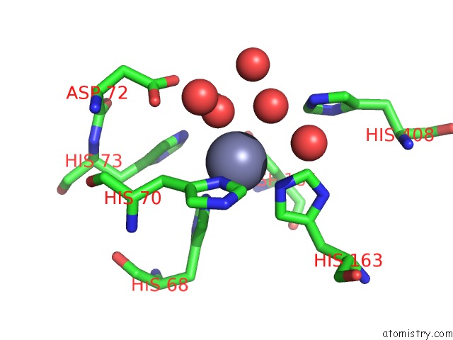
Mono view
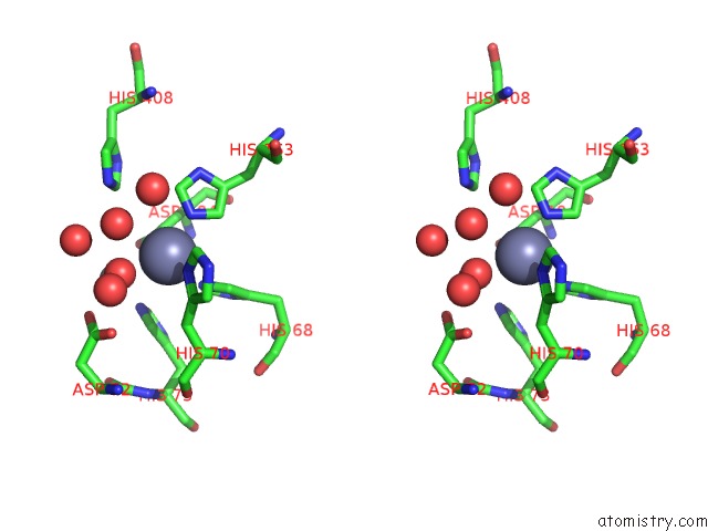
Stereo pair view
Mono view
Stereo pair view
A full contact list of Zinc with other atoms in the Zn binding
site number 2 of Structure of the YSH1-MPE1 Nuclease Complex From S.Cerevisiae within 5.0Å range:
|
Reference:
C.H.Hill,
V.Boreikaite,
A.Kumar,
A.Casanal,
P.Kubik,
G.Degliesposti,
S.Maslen,
A.Mariani,
O.Von Loeffelholz,
M.Girbig,
M.Skehel,
L.A.Passmore.
Activation of the Endonuclease That Defines Mrna 3' Ends Requires Incorporation Into An 8-Subunit Core Cleavage and Polyadenylation Factor Complex. Mol.Cell V. 73 1217 2019.
ISSN: ISSN 1097-2765
PubMed: 30737185
DOI: 10.1016/J.MOLCEL.2018.12.023
Page generated: Mon Oct 28 23:34:40 2024
ISSN: ISSN 1097-2765
PubMed: 30737185
DOI: 10.1016/J.MOLCEL.2018.12.023
Last articles
Zn in 9MJ5Zn in 9HNW
Zn in 9G0L
Zn in 9FNE
Zn in 9DZN
Zn in 9E0I
Zn in 9D32
Zn in 9DAK
Zn in 8ZXC
Zn in 8ZUF