Zinc »
PDB 6h56-6hcz »
6h5w »
Zinc in PDB 6h5w: Crystal Structure of Human Angiotensin-1 Converting Enzyme C-Domain in Complex with Omapatrilat.
Enzymatic activity of Crystal Structure of Human Angiotensin-1 Converting Enzyme C-Domain in Complex with Omapatrilat.
All present enzymatic activity of Crystal Structure of Human Angiotensin-1 Converting Enzyme C-Domain in Complex with Omapatrilat.:
3.4.15.1;
3.4.15.1;
Protein crystallography data
The structure of Crystal Structure of Human Angiotensin-1 Converting Enzyme C-Domain in Complex with Omapatrilat., PDB code: 6h5w
was solved by
G.E.Cozier,
K.R.Acharya,
with X-Ray Crystallography technique. A brief refinement statistics is given in the table below:
| Resolution Low / High (Å) | 71.89 / 1.37 |
| Space group | P 21 21 21 |
| Cell size a, b, c (Å), α, β, γ (°) | 56.596, 85.126, 134.264, 90.00, 90.00, 90.00 |
| R / Rfree (%) | 14.5 / 17.5 |
Other elements in 6h5w:
The structure of Crystal Structure of Human Angiotensin-1 Converting Enzyme C-Domain in Complex with Omapatrilat. also contains other interesting chemical elements:
| Chlorine | (Cl) | 2 atoms |
Zinc Binding Sites:
The binding sites of Zinc atom in the Crystal Structure of Human Angiotensin-1 Converting Enzyme C-Domain in Complex with Omapatrilat.
(pdb code 6h5w). This binding sites where shown within
5.0 Angstroms radius around Zinc atom.
In total only one binding site of Zinc was determined in the Crystal Structure of Human Angiotensin-1 Converting Enzyme C-Domain in Complex with Omapatrilat., PDB code: 6h5w:
In total only one binding site of Zinc was determined in the Crystal Structure of Human Angiotensin-1 Converting Enzyme C-Domain in Complex with Omapatrilat., PDB code: 6h5w:
Zinc binding site 1 out of 1 in 6h5w
Go back to
Zinc binding site 1 out
of 1 in the Crystal Structure of Human Angiotensin-1 Converting Enzyme C-Domain in Complex with Omapatrilat.
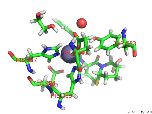
Mono view
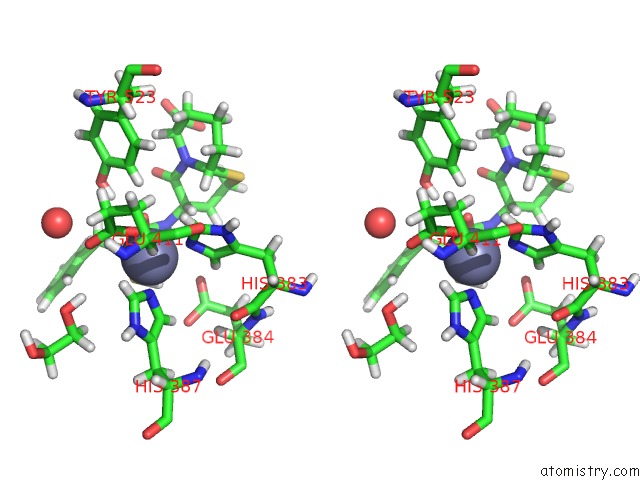
Stereo pair view
Mono view
Stereo pair view
A full contact list of Zinc with other atoms in the Zn binding
site number 1 of Crystal Structure of Human Angiotensin-1 Converting Enzyme C-Domain in Complex with Omapatrilat. within 5.0Å range:
|
Reference:
G.E.Cozier,
L.B.Arendse,
S.L.Schwager,
E.D.Sturrock,
K.R.Acharya.
Molecular Basis For Multiple Omapatrilat Binding Sites Within the Ace C-Domain: Implications For Drug Design. J. Med. Chem. V. 61 10141 2018.
ISSN: ISSN 1520-4804
PubMed: 30372620
DOI: 10.1021/ACS.JMEDCHEM.8B01309
Page generated: Mon Oct 28 22:37:20 2024
ISSN: ISSN 1520-4804
PubMed: 30372620
DOI: 10.1021/ACS.JMEDCHEM.8B01309
Last articles
Zn in 9MJ5Zn in 9HNW
Zn in 9G0L
Zn in 9FNE
Zn in 9DZN
Zn in 9E0I
Zn in 9D32
Zn in 9DAK
Zn in 8ZXC
Zn in 8ZUF