Zinc »
PDB 6fso-6g3m »
6fuk »
Zinc in PDB 6fuk: Crystal Structure of Utx Complexed with 5-Carboxy-8-Hydroxyquinoline
Protein crystallography data
The structure of Crystal Structure of Utx Complexed with 5-Carboxy-8-Hydroxyquinoline, PDB code: 6fuk
was solved by
C.Esposito,
P.Sledz,
A.Caflisch,
with X-Ray Crystallography technique. A brief refinement statistics is given in the table below:
| Resolution Low / High (Å) | 46.45 / 2.00 |
| Space group | P 21 21 21 |
| Cell size a, b, c (Å), α, β, γ (°) | 80.417, 83.261, 92.898, 90.00, 90.00, 90.00 |
| R / Rfree (%) | 18.6 / 22.8 |
Other elements in 6fuk:
The structure of Crystal Structure of Utx Complexed with 5-Carboxy-8-Hydroxyquinoline also contains other interesting chemical elements:
| Manganese | (Mn) | 1 atom |
Zinc Binding Sites:
The binding sites of Zinc atom in the Crystal Structure of Utx Complexed with 5-Carboxy-8-Hydroxyquinoline
(pdb code 6fuk). This binding sites where shown within
5.0 Angstroms radius around Zinc atom.
In total only one binding site of Zinc was determined in the Crystal Structure of Utx Complexed with 5-Carboxy-8-Hydroxyquinoline, PDB code: 6fuk:
In total only one binding site of Zinc was determined in the Crystal Structure of Utx Complexed with 5-Carboxy-8-Hydroxyquinoline, PDB code: 6fuk:
Zinc binding site 1 out of 1 in 6fuk
Go back to
Zinc binding site 1 out
of 1 in the Crystal Structure of Utx Complexed with 5-Carboxy-8-Hydroxyquinoline
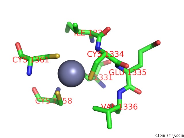
Mono view
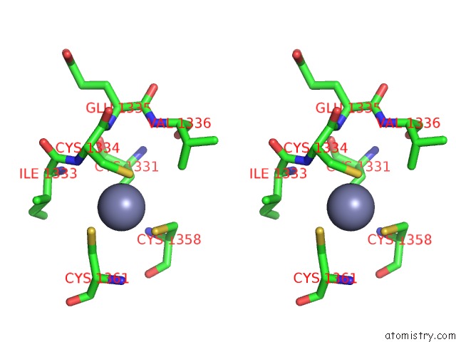
Stereo pair view
Mono view
Stereo pair view
A full contact list of Zinc with other atoms in the Zn binding
site number 1 of Crystal Structure of Utx Complexed with 5-Carboxy-8-Hydroxyquinoline within 5.0Å range:
|
Reference:
C.Esposito,
L.Wiedmer,
A.Caflisch.
In Silico Identification of JMJD3 Demethylase Inhibitors. J Chem Inf Model V. 58 2151 2018.
ISSN: ESSN 1549-960X
PubMed: 30226987
DOI: 10.1021/ACS.JCIM.8B00539
Page generated: Mon Oct 28 21:25:36 2024
ISSN: ESSN 1549-960X
PubMed: 30226987
DOI: 10.1021/ACS.JCIM.8B00539
Last articles
Zn in 9MJ5Zn in 9HNW
Zn in 9G0L
Zn in 9FNE
Zn in 9DZN
Zn in 9E0I
Zn in 9D32
Zn in 9DAK
Zn in 8ZXC
Zn in 8ZUF