Zinc »
PDB 6fhh-6fsm »
6fjj »
Zinc in PDB 6fjj: Joint Neutron and X-Ray Crystal Structure of Human Carbonic Anhydrase IX Mimic (Saccharin).
Enzymatic activity of Joint Neutron and X-Ray Crystal Structure of Human Carbonic Anhydrase IX Mimic (Saccharin).
All present enzymatic activity of Joint Neutron and X-Ray Crystal Structure of Human Carbonic Anhydrase IX Mimic (Saccharin).:
4.2.1.1;
4.2.1.1;
Protein crystallography data
The structure of Joint Neutron and X-Ray Crystal Structure of Human Carbonic Anhydrase IX Mimic (Saccharin)., PDB code: 6fjj
was solved by
S.Z.Fisher,
with X-Ray Crystallography technique. A brief refinement statistics is given in the table below:
| Resolution Low / High (Å) | N/A / 1.50 |
| Space group | P 1 21 1 |
| Cell size a, b, c (Å), α, β, γ (°) | 42.530, 41.830, 72.700, 90.00, 104.04, 90.00 |
| R / Rfree (%) | 20.5 / 23.1 |
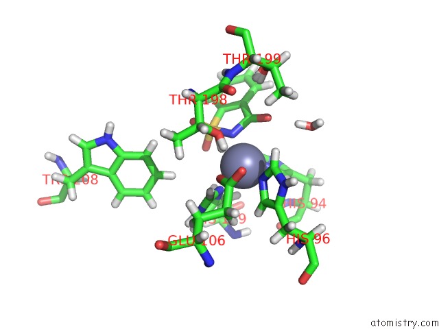
Zinc Binding Sites:
The binding sites of Zinc atom in the Joint Neutron and X-Ray Crystal Structure of Human Carbonic Anhydrase IX Mimic (Saccharin).
(pdb code 6fjj). This binding sites where shown within
5.0 Angstroms radius around Zinc atom.
In total only one binding site of Zinc was determined in the Joint Neutron and X-Ray Crystal Structure of Human Carbonic Anhydrase IX Mimic (Saccharin)., PDB code: 6fjj:
In total only one binding site of Zinc was determined in the Joint Neutron and X-Ray Crystal Structure of Human Carbonic Anhydrase IX Mimic (Saccharin)., PDB code: 6fjj:
Zinc binding site 1 out of 1 in 6fjj
Go back to
Zinc binding site 1 out
of 1 in the Joint Neutron and X-Ray Crystal Structure of Human Carbonic Anhydrase IX Mimic (Saccharin).
Mono view
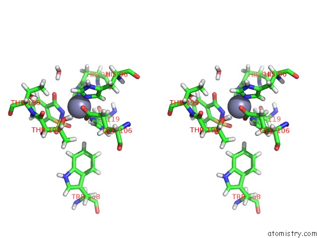
Stereo pair view
Mono view
Stereo pair view
A full contact list of Zinc with other atoms in the Zn binding
site number 1 of Joint Neutron and X-Ray Crystal Structure of Human Carbonic Anhydrase IX Mimic (Saccharin). within 5.0Å range:
|
Reference:
K.Koruza,
B.P.Mahon,
M.P.Blakeley,
A.Ostermann,
T.E.Schrader,
R.Mckenna,
W.Knecht,
S.Z.Fisher.
Using Neutron Crystallography to Elucidate the Basis of Selective Inhibition of Carbonic Anhydrase By Saccharin and A Derivative. J. Struct. Biol. V. 205 147 2019.
ISSN: ESSN 1095-8657
PubMed: 30639924
DOI: 10.1016/J.JSB.2018.12.009
Page generated: Mon Oct 28 21:07:45 2024
ISSN: ESSN 1095-8657
PubMed: 30639924
DOI: 10.1016/J.JSB.2018.12.009
Last articles
Zn in 9MJ5Zn in 9HNW
Zn in 9G0L
Zn in 9FNE
Zn in 9DZN
Zn in 9E0I
Zn in 9D32
Zn in 9DAK
Zn in 8ZXC
Zn in 8ZUF