Zinc »
PDB 6f9r-6fhg »
6fbq »
Zinc in PDB 6fbq: Crystal Structure of the Human Retinoid X Receptor Dna-Binding Domain Bound to the Human Mep DR1 Response Element, pH 7.0
Protein crystallography data
The structure of Crystal Structure of the Human Retinoid X Receptor Dna-Binding Domain Bound to the Human Mep DR1 Response Element, pH 7.0, PDB code: 6fbq
was solved by
A.G.Mcewen,
P.Poussin-Courmontagne,
J.Osz,
N.Rochel,
with X-Ray Crystallography technique. A brief refinement statistics is given in the table below:
| Resolution Low / High (Å) | 42.44 / 1.60 |
| Space group | C 1 2 1 |
| Cell size a, b, c (Å), α, β, γ (°) | 103.333, 44.257, 62.137, 90.00, 98.31, 90.00 |
| R / Rfree (%) | 16.3 / 19.8 |
Zinc Binding Sites:
The binding sites of Zinc atom in the Crystal Structure of the Human Retinoid X Receptor Dna-Binding Domain Bound to the Human Mep DR1 Response Element, pH 7.0
(pdb code 6fbq). This binding sites where shown within
5.0 Angstroms radius around Zinc atom.
In total 4 binding sites of Zinc where determined in the Crystal Structure of the Human Retinoid X Receptor Dna-Binding Domain Bound to the Human Mep DR1 Response Element, pH 7.0, PDB code: 6fbq:
Jump to Zinc binding site number: 1; 2; 3; 4;
In total 4 binding sites of Zinc where determined in the Crystal Structure of the Human Retinoid X Receptor Dna-Binding Domain Bound to the Human Mep DR1 Response Element, pH 7.0, PDB code: 6fbq:
Jump to Zinc binding site number: 1; 2; 3; 4;
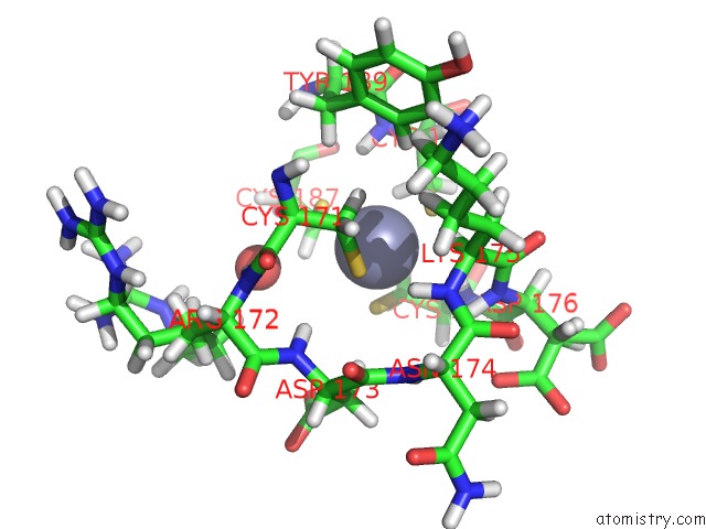
Zinc binding site 1 out of 4 in 6fbq
Go back to
Zinc binding site 1 out
of 4 in the Crystal Structure of the Human Retinoid X Receptor Dna-Binding Domain Bound to the Human Mep DR1 Response Element, pH 7.0
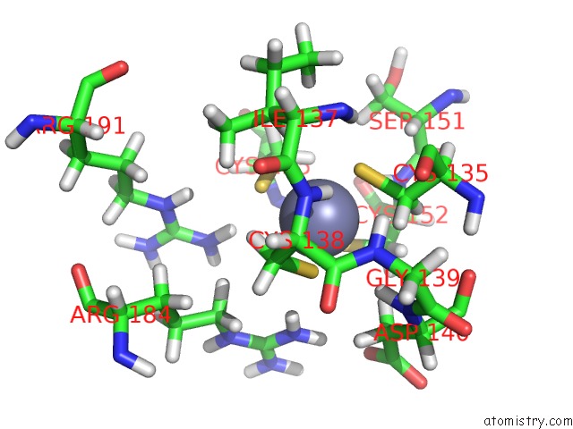
Mono view
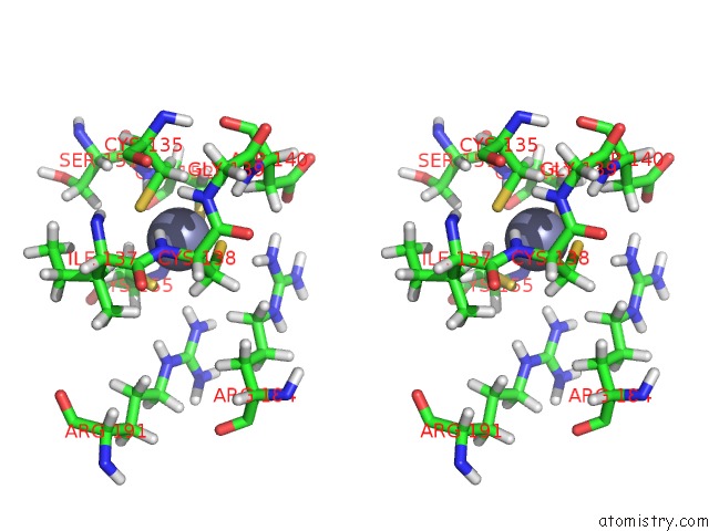
Stereo pair view
Mono view
Stereo pair view
A full contact list of Zinc with other atoms in the Zn binding
site number 1 of Crystal Structure of the Human Retinoid X Receptor Dna-Binding Domain Bound to the Human Mep DR1 Response Element, pH 7.0 within 5.0Å range:
|
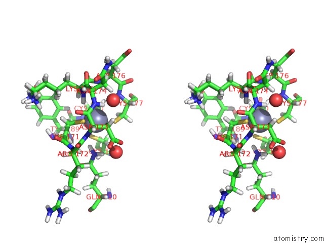
Zinc binding site 2 out of 4 in 6fbq
Go back to
Zinc binding site 2 out
of 4 in the Crystal Structure of the Human Retinoid X Receptor Dna-Binding Domain Bound to the Human Mep DR1 Response Element, pH 7.0

Mono view
Stereo pair view
Mono view
Stereo pair view
A full contact list of Zinc with other atoms in the Zn binding
site number 2 of Crystal Structure of the Human Retinoid X Receptor Dna-Binding Domain Bound to the Human Mep DR1 Response Element, pH 7.0 within 5.0Å range:
|
Zinc binding site 3 out of 4 in 6fbq
Go back to
Zinc binding site 3 out
of 4 in the Crystal Structure of the Human Retinoid X Receptor Dna-Binding Domain Bound to the Human Mep DR1 Response Element, pH 7.0
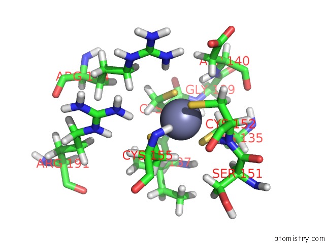
Mono view

Stereo pair view
Mono view
Stereo pair view
A full contact list of Zinc with other atoms in the Zn binding
site number 3 of Crystal Structure of the Human Retinoid X Receptor Dna-Binding Domain Bound to the Human Mep DR1 Response Element, pH 7.0 within 5.0Å range:
|
Zinc binding site 4 out of 4 in 6fbq
Go back to
Zinc binding site 4 out
of 4 in the Crystal Structure of the Human Retinoid X Receptor Dna-Binding Domain Bound to the Human Mep DR1 Response Element, pH 7.0
Mono view

Stereo pair view
Mono view
Stereo pair view
A full contact list of Zinc with other atoms in the Zn binding
site number 4 of Crystal Structure of the Human Retinoid X Receptor Dna-Binding Domain Bound to the Human Mep DR1 Response Element, pH 7.0 within 5.0Å range:
|
Reference:
J.Osz,
A.G.Mcewen,
J.Wolf,
P.Poussin-Courmontagne,
C.Peluso-Iltis,
Y.Chebaro,
B.Kieffer,
N.Rochel.
Modulation of Rxr-Dna Complex Assembly By Dna Context. Mol. Cell. Endocrinol. V. 481 44 2019.
ISSN: ISSN 1872-8057
PubMed: 30476562
DOI: 10.1016/J.MCE.2018.11.008
Page generated: Mon Oct 28 20:49:29 2024
ISSN: ISSN 1872-8057
PubMed: 30476562
DOI: 10.1016/J.MCE.2018.11.008
Last articles
Zn in 9MJ5Zn in 9HNW
Zn in 9G0L
Zn in 9FNE
Zn in 9DZN
Zn in 9E0I
Zn in 9D32
Zn in 9DAK
Zn in 8ZXC
Zn in 8ZUF