Zinc »
PDB 6eea-6eox »
6egl »
Zinc in PDB 6egl: Crystal Structure of A De Novo Three-Stranded Coiled Coil Peptide Containing A D-Leu in the Second Coordination Sphere of A Non- Metalated Tris-Thiolate Binding Site
Protein crystallography data
The structure of Crystal Structure of A De Novo Three-Stranded Coiled Coil Peptide Containing A D-Leu in the Second Coordination Sphere of A Non- Metalated Tris-Thiolate Binding Site, PDB code: 6egl
was solved by
L.Ruckthong,
J.A.Stuckey,
V.L.Pecoraro,
with X-Ray Crystallography technique. A brief refinement statistics is given in the table below:
| Resolution Low / High (Å) | 9.28 / 1.40 |
| Space group | H 3 2 |
| Cell size a, b, c (Å), α, β, γ (°) | 38.213, 38.213, 140.655, 90.00, 90.00, 120.00 |
| R / Rfree (%) | 17.7 / 18.4 |
Zinc Binding Sites:
The binding sites of Zinc atom in the Crystal Structure of A De Novo Three-Stranded Coiled Coil Peptide Containing A D-Leu in the Second Coordination Sphere of A Non- Metalated Tris-Thiolate Binding Site
(pdb code 6egl). This binding sites where shown within
5.0 Angstroms radius around Zinc atom.
In total only one binding site of Zinc was determined in the Crystal Structure of A De Novo Three-Stranded Coiled Coil Peptide Containing A D-Leu in the Second Coordination Sphere of A Non- Metalated Tris-Thiolate Binding Site, PDB code: 6egl:
In total only one binding site of Zinc was determined in the Crystal Structure of A De Novo Three-Stranded Coiled Coil Peptide Containing A D-Leu in the Second Coordination Sphere of A Non- Metalated Tris-Thiolate Binding Site, PDB code: 6egl:
Zinc binding site 1 out of 1 in 6egl
Go back to
Zinc binding site 1 out
of 1 in the Crystal Structure of A De Novo Three-Stranded Coiled Coil Peptide Containing A D-Leu in the Second Coordination Sphere of A Non- Metalated Tris-Thiolate Binding Site
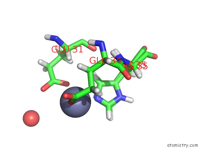
Mono view
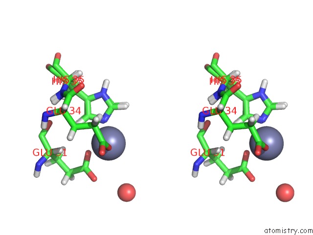
Stereo pair view
Mono view
Stereo pair view
A full contact list of Zinc with other atoms in the Zn binding
site number 1 of Crystal Structure of A De Novo Three-Stranded Coiled Coil Peptide Containing A D-Leu in the Second Coordination Sphere of A Non- Metalated Tris-Thiolate Binding Site within 5.0Å range:
|
Reference:
L.Ruckthong,
J.A.Stuckey,
V.L.Pecoraro.
How Outer Coordination Sphere Modifications Can Impact Metal Structures in Proteins: A Crystallographic Evaluation. Chemistry V. 25 6773 2019.
ISSN: ISSN 0947-6539
PubMed: 30861211
DOI: 10.1002/CHEM.201806040
Page generated: Mon Oct 28 20:08:25 2024
ISSN: ISSN 0947-6539
PubMed: 30861211
DOI: 10.1002/CHEM.201806040
Last articles
Zn in 9MJ5Zn in 9HNW
Zn in 9G0L
Zn in 9FNE
Zn in 9DZN
Zn in 9E0I
Zn in 9D32
Zn in 9DAK
Zn in 8ZXC
Zn in 8ZUF