Zinc »
PDB 6eea-6eox »
6eee »
Zinc in PDB 6eee: X-Ray Crystal Structure of Pf-M17 in Complex with Inhibitor (6K) and Regulatory Zinc Ion
Protein crystallography data
The structure of X-Ray Crystal Structure of Pf-M17 in Complex with Inhibitor (6K) and Regulatory Zinc Ion, PDB code: 6eee
was solved by
N.Drinkwater,
S.Mcgowan,
with X-Ray Crystallography technique. A brief refinement statistics is given in the table below:
| Resolution Low / High (Å) | 48.17 / 2.30 |
| Space group | P 21 21 21 |
| Cell size a, b, c (Å), α, β, γ (°) | 173.680, 177.395, 229.445, 90.00, 90.00, 90.00 |
| R / Rfree (%) | 19.3 / 24.6 |
Other elements in 6eee:
The structure of X-Ray Crystal Structure of Pf-M17 in Complex with Inhibitor (6K) and Regulatory Zinc Ion also contains other interesting chemical elements:
| Fluorine | (F) | 36 atoms |
Zinc Binding Sites:
Pages:
>>> Page 1 <<< Page 2, Binding sites: 11 - 12;Binding sites:
The binding sites of Zinc atom in the X-Ray Crystal Structure of Pf-M17 in Complex with Inhibitor (6K) and Regulatory Zinc Ion (pdb code 6eee). This binding sites where shown within 5.0 Angstroms radius around Zinc atom.In total 12 binding sites of Zinc where determined in the X-Ray Crystal Structure of Pf-M17 in Complex with Inhibitor (6K) and Regulatory Zinc Ion, PDB code: 6eee:
Jump to Zinc binding site number: 1; 2; 3; 4; 5; 6; 7; 8; 9; 10;
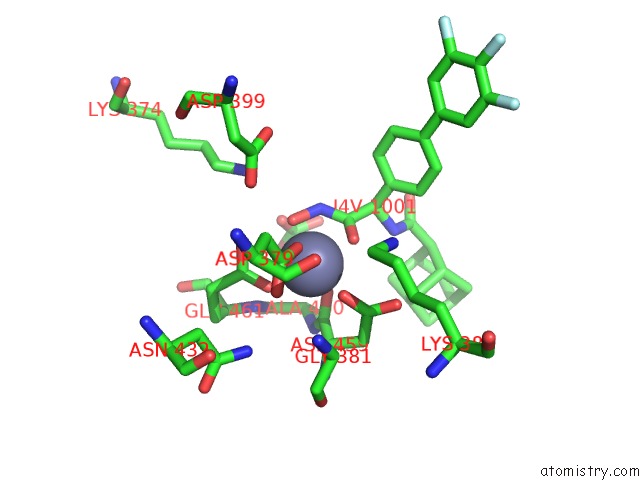
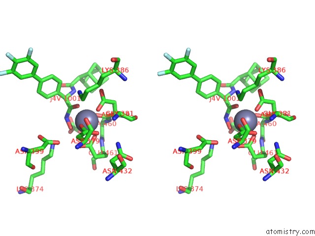
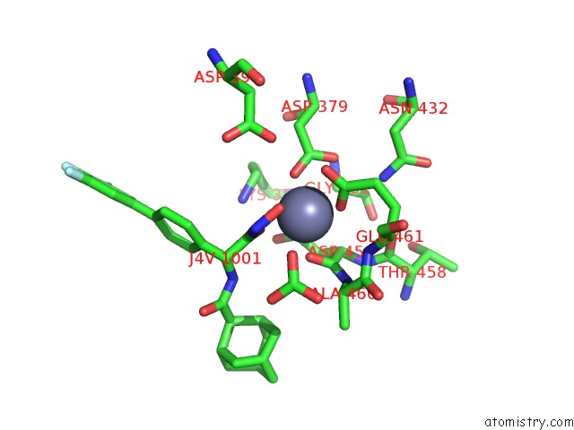
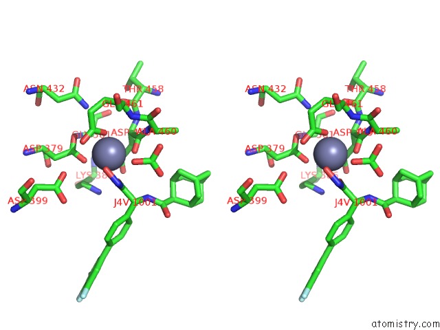
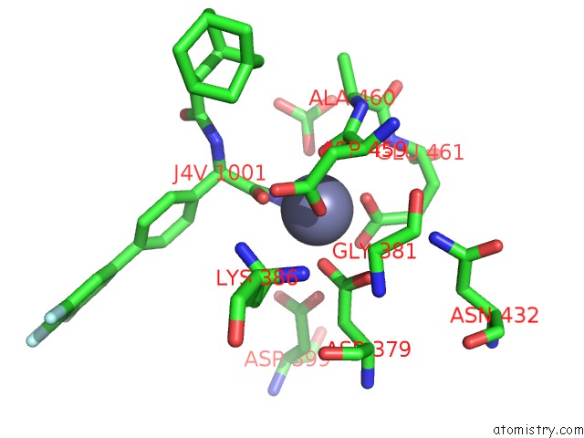
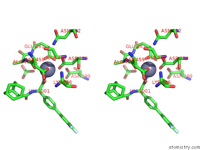
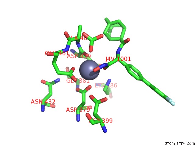
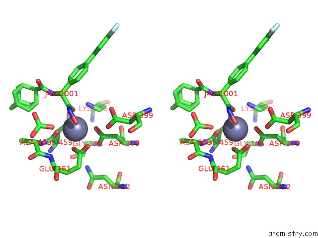
Zinc binding site 1 out of 12 in 6eee
Go back to
Zinc binding site 1 out
of 12 in the X-Ray Crystal Structure of Pf-M17 in Complex with Inhibitor (6K) and Regulatory Zinc Ion
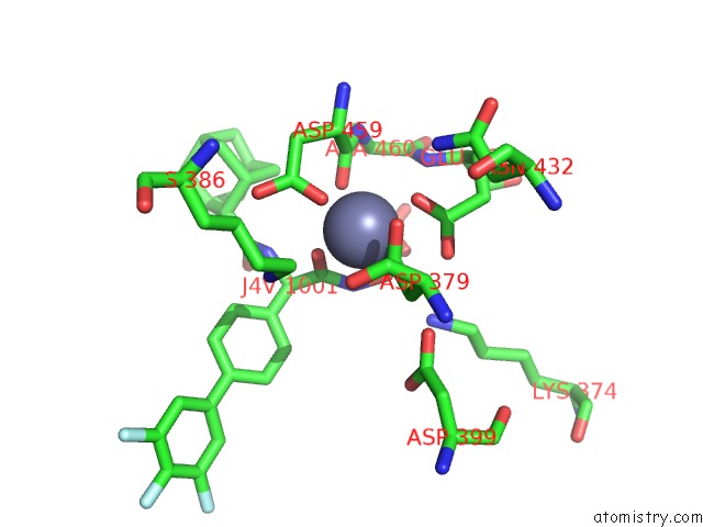
Mono view
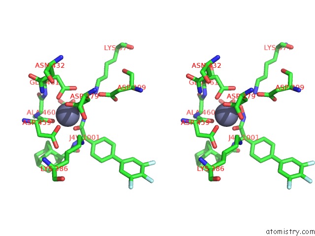
Stereo pair view
Mono view
Stereo pair view
A full contact list of Zinc with other atoms in the Zn binding
site number 1 of X-Ray Crystal Structure of Pf-M17 in Complex with Inhibitor (6K) and Regulatory Zinc Ion within 5.0Å range:
|
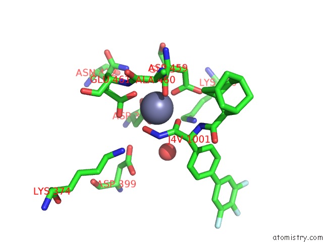
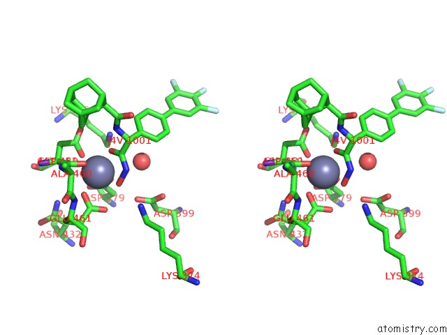
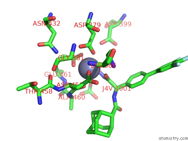
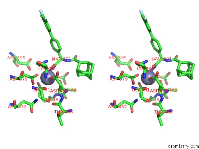
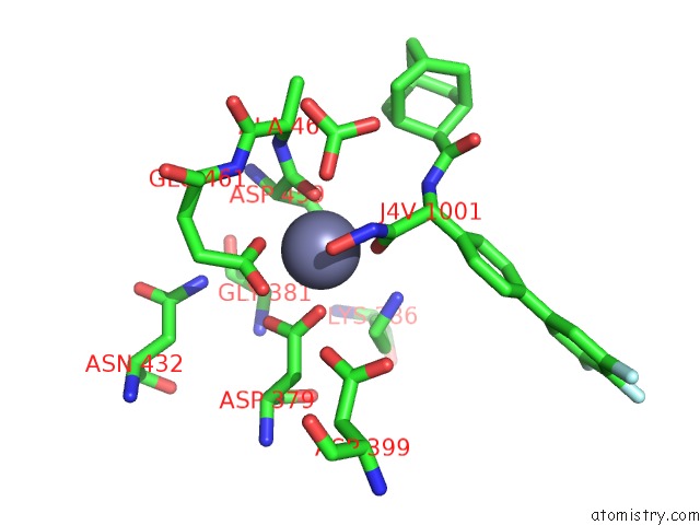
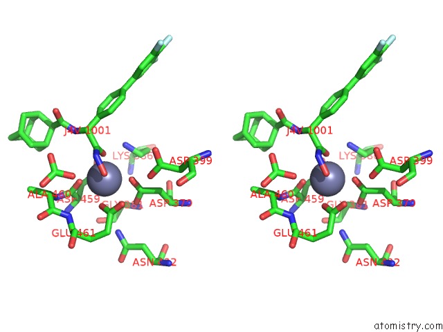
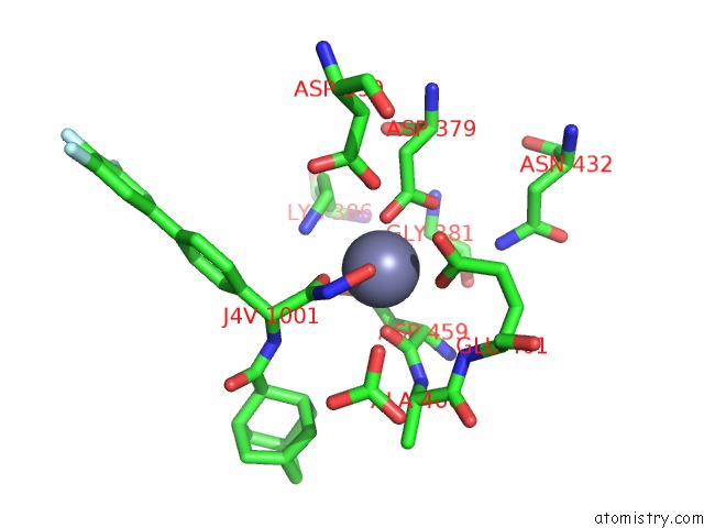
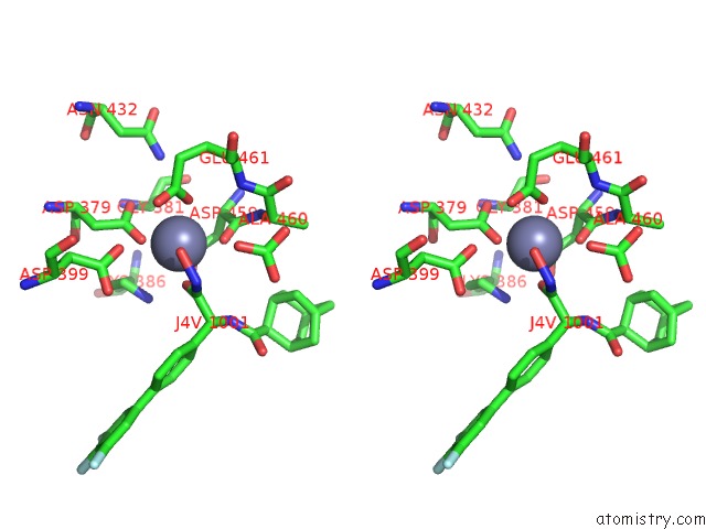
Zinc binding site 2 out of 12 in 6eee
Go back to
Zinc binding site 2 out
of 12 in the X-Ray Crystal Structure of Pf-M17 in Complex with Inhibitor (6K) and Regulatory Zinc Ion
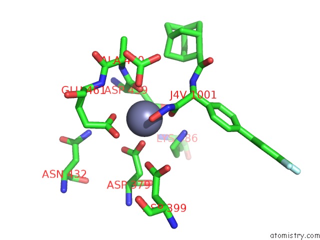
Mono view
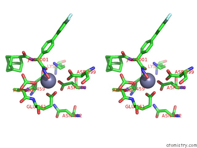
Stereo pair view
Mono view
Stereo pair view
A full contact list of Zinc with other atoms in the Zn binding
site number 2 of X-Ray Crystal Structure of Pf-M17 in Complex with Inhibitor (6K) and Regulatory Zinc Ion within 5.0Å range:
|
Zinc binding site 3 out of 12 in 6eee
Go back to
Zinc binding site 3 out
of 12 in the X-Ray Crystal Structure of Pf-M17 in Complex with Inhibitor (6K) and Regulatory Zinc Ion
Mono view
Stereo pair view
Mono view
Stereo pair view
A full contact list of Zinc with other atoms in the Zn binding
site number 3 of X-Ray Crystal Structure of Pf-M17 in Complex with Inhibitor (6K) and Regulatory Zinc Ion within 5.0Å range:
|
Zinc binding site 4 out of 12 in 6eee
Go back to
Zinc binding site 4 out
of 12 in the X-Ray Crystal Structure of Pf-M17 in Complex with Inhibitor (6K) and Regulatory Zinc Ion
Mono view
Stereo pair view
Mono view
Stereo pair view
A full contact list of Zinc with other atoms in the Zn binding
site number 4 of X-Ray Crystal Structure of Pf-M17 in Complex with Inhibitor (6K) and Regulatory Zinc Ion within 5.0Å range:
|
Zinc binding site 5 out of 12 in 6eee
Go back to
Zinc binding site 5 out
of 12 in the X-Ray Crystal Structure of Pf-M17 in Complex with Inhibitor (6K) and Regulatory Zinc Ion
Mono view
Stereo pair view
Mono view
Stereo pair view
A full contact list of Zinc with other atoms in the Zn binding
site number 5 of X-Ray Crystal Structure of Pf-M17 in Complex with Inhibitor (6K) and Regulatory Zinc Ion within 5.0Å range:
|
Zinc binding site 6 out of 12 in 6eee
Go back to
Zinc binding site 6 out
of 12 in the X-Ray Crystal Structure of Pf-M17 in Complex with Inhibitor (6K) and Regulatory Zinc Ion
Mono view
Stereo pair view
Mono view
Stereo pair view
A full contact list of Zinc with other atoms in the Zn binding
site number 6 of X-Ray Crystal Structure of Pf-M17 in Complex with Inhibitor (6K) and Regulatory Zinc Ion within 5.0Å range:
|
Zinc binding site 7 out of 12 in 6eee
Go back to
Zinc binding site 7 out
of 12 in the X-Ray Crystal Structure of Pf-M17 in Complex with Inhibitor (6K) and Regulatory Zinc Ion
Mono view
Stereo pair view
Mono view
Stereo pair view
A full contact list of Zinc with other atoms in the Zn binding
site number 7 of X-Ray Crystal Structure of Pf-M17 in Complex with Inhibitor (6K) and Regulatory Zinc Ion within 5.0Å range:
|
Zinc binding site 8 out of 12 in 6eee
Go back to
Zinc binding site 8 out
of 12 in the X-Ray Crystal Structure of Pf-M17 in Complex with Inhibitor (6K) and Regulatory Zinc Ion
Mono view
Stereo pair view
Mono view
Stereo pair view
A full contact list of Zinc with other atoms in the Zn binding
site number 8 of X-Ray Crystal Structure of Pf-M17 in Complex with Inhibitor (6K) and Regulatory Zinc Ion within 5.0Å range:
|
Zinc binding site 9 out of 12 in 6eee
Go back to
Zinc binding site 9 out
of 12 in the X-Ray Crystal Structure of Pf-M17 in Complex with Inhibitor (6K) and Regulatory Zinc Ion
Mono view
Stereo pair view
Mono view
Stereo pair view
A full contact list of Zinc with other atoms in the Zn binding
site number 9 of X-Ray Crystal Structure of Pf-M17 in Complex with Inhibitor (6K) and Regulatory Zinc Ion within 5.0Å range:
|
Zinc binding site 10 out of 12 in 6eee
Go back to
Zinc binding site 10 out
of 12 in the X-Ray Crystal Structure of Pf-M17 in Complex with Inhibitor (6K) and Regulatory Zinc Ion
Mono view
Stereo pair view
Mono view
Stereo pair view
A full contact list of Zinc with other atoms in the Zn binding
site number 10 of X-Ray Crystal Structure of Pf-M17 in Complex with Inhibitor (6K) and Regulatory Zinc Ion within 5.0Å range:
|
Reference:
N.B.Vinh,
N.Drinkwater,
T.R.Malcolm,
M.Kassiou,
L.Lucantoni,
P.M.Grin,
G.S.Butler,
S.Duffy,
C.M.Overall,
V.M.Avery,
P.J.Scammells,
S.Mcgowan.
Hydroxamic Acid Inhibitors Provide Cross-Species Inhibition of Plasmodium M1 and M17 Aminopeptidases. J. Med. Chem. V. 62 622 2019.
ISSN: ISSN 1520-4804
PubMed: 30537832
DOI: 10.1021/ACS.JMEDCHEM.8B01310
Page generated: Mon Oct 28 20:07:57 2024
ISSN: ISSN 1520-4804
PubMed: 30537832
DOI: 10.1021/ACS.JMEDCHEM.8B01310
Last articles
Zn in 9MJ5Zn in 9HNW
Zn in 9G0L
Zn in 9FNE
Zn in 9DZN
Zn in 9E0I
Zn in 9D32
Zn in 9DAK
Zn in 8ZXC
Zn in 8ZUF