Zinc »
PDB 6dy5-6ee8 »
6dz8 »
Zinc in PDB 6dz8: Crystal Structure of S. Aureus Penicillin Binding Protein 4 (PBP4) Mutant (S75C)
Protein crystallography data
The structure of Crystal Structure of S. Aureus Penicillin Binding Protein 4 (PBP4) Mutant (S75C), PDB code: 6dz8
was solved by
J.A.N.Alexander,
N.C.J.Strynadka,
with X-Ray Crystallography technique. A brief refinement statistics is given in the table below:
| Resolution Low / High (Å) | 29.16 / 1.86 |
| Space group | C 1 2 1 |
| Cell size a, b, c (Å), α, β, γ (°) | 104.511, 89.730, 77.489, 90.00, 98.09, 90.00 |
| R / Rfree (%) | 17.5 / 21.7 |
Zinc Binding Sites:
The binding sites of Zinc atom in the Crystal Structure of S. Aureus Penicillin Binding Protein 4 (PBP4) Mutant (S75C)
(pdb code 6dz8). This binding sites where shown within
5.0 Angstroms radius around Zinc atom.
In total 4 binding sites of Zinc where determined in the Crystal Structure of S. Aureus Penicillin Binding Protein 4 (PBP4) Mutant (S75C), PDB code: 6dz8:
Jump to Zinc binding site number: 1; 2; 3; 4;
In total 4 binding sites of Zinc where determined in the Crystal Structure of S. Aureus Penicillin Binding Protein 4 (PBP4) Mutant (S75C), PDB code: 6dz8:
Jump to Zinc binding site number: 1; 2; 3; 4;
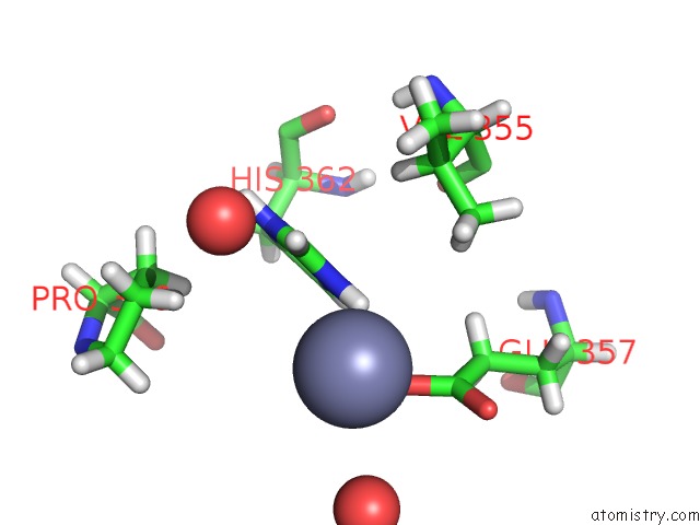
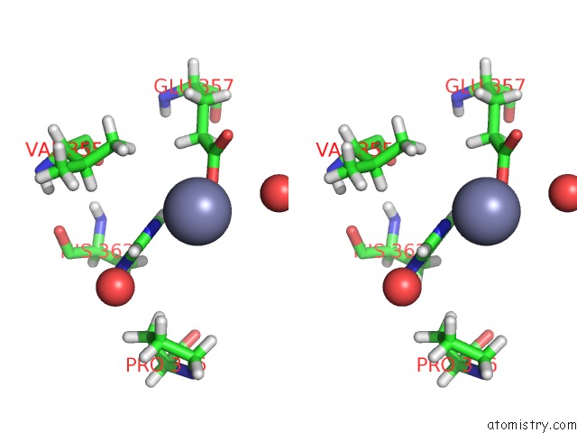
Zinc binding site 1 out of 4 in 6dz8
Go back to
Zinc binding site 1 out
of 4 in the Crystal Structure of S. Aureus Penicillin Binding Protein 4 (PBP4) Mutant (S75C)
Mono view

Stereo pair view
Mono view
Stereo pair view
A full contact list of Zinc with other atoms in the Zn binding
site number 1 of Crystal Structure of S. Aureus Penicillin Binding Protein 4 (PBP4) Mutant (S75C) within 5.0Å range:
|
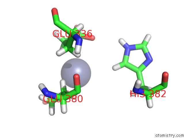
Zinc binding site 2 out of 4 in 6dz8
Go back to
Zinc binding site 2 out
of 4 in the Crystal Structure of S. Aureus Penicillin Binding Protein 4 (PBP4) Mutant (S75C)
Mono view

Stereo pair view
Mono view
Stereo pair view
A full contact list of Zinc with other atoms in the Zn binding
site number 2 of Crystal Structure of S. Aureus Penicillin Binding Protein 4 (PBP4) Mutant (S75C) within 5.0Å range:
|
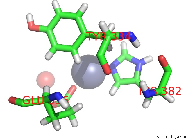
Zinc binding site 3 out of 4 in 6dz8
Go back to
Zinc binding site 3 out
of 4 in the Crystal Structure of S. Aureus Penicillin Binding Protein 4 (PBP4) Mutant (S75C)
Mono view

Stereo pair view
Mono view
Stereo pair view
A full contact list of Zinc with other atoms in the Zn binding
site number 3 of Crystal Structure of S. Aureus Penicillin Binding Protein 4 (PBP4) Mutant (S75C) within 5.0Å range:
|
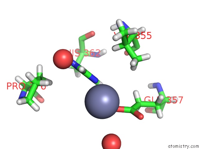
Zinc binding site 4 out of 4 in 6dz8
Go back to
Zinc binding site 4 out
of 4 in the Crystal Structure of S. Aureus Penicillin Binding Protein 4 (PBP4) Mutant (S75C)
Mono view
Stereo pair view
Mono view
Stereo pair view
A full contact list of Zinc with other atoms in the Zn binding
site number 4 of Crystal Structure of S. Aureus Penicillin Binding Protein 4 (PBP4) Mutant (S75C) within 5.0Å range:
|
Reference:
R.Maya-Martinez,
J.A.N.Alexander,
C.F.Otten,
I.Ayala,
D.Vollmer,
J.Gray,
C.M.Bougault,
A.Burt,
C.Laguri,
M.Fonvielle,
M.Arthur,
N.C.J.Strynadka,
W.Vollmer,
J.P.Simorre.
Recognition of Peptidoglycan Fragments By the Transpeptidase PBP4 Fromstaphylococcus Aureus. Front Microbiol V. 9 3223 2018.
ISSN: ESSN 1664-302X
PubMed: 30713527
DOI: 10.3389/FMICB.2018.03223
Page generated: Mon Oct 28 19:56:21 2024
ISSN: ESSN 1664-302X
PubMed: 30713527
DOI: 10.3389/FMICB.2018.03223
Last articles
Zn in 9MJ5Zn in 9HNW
Zn in 9G0L
Zn in 9FNE
Zn in 9DZN
Zn in 9E0I
Zn in 9D32
Zn in 9DAK
Zn in 8ZXC
Zn in 8ZUF