Zinc in PDB 6djx: Crystal Structure of Pparkin-Pub-UBCH7 Complex
Enzymatic activity of Crystal Structure of Pparkin-Pub-UBCH7 Complex
All present enzymatic activity of Crystal Structure of Pparkin-Pub-UBCH7 Complex:
2.3.2.23;
2.3.2.31;
Protein crystallography data
The structure of Crystal Structure of Pparkin-Pub-UBCH7 Complex, PDB code: 6djx
was solved by
V.Sauve,
G.Sung,
J.F.Trempe,
K.Gehring,
with X-Ray Crystallography technique. A brief refinement statistics is given in the table below:
Resolution Low / High (Å)
|
48.86 /
4.80
|
Space group
|
P 31 2 1
|
Cell size a, b, c (Å), α, β, γ (°)
|
135.669,
135.669,
87.993,
90.00,
90.00,
120.00
|
R / Rfree (%)
|
25.7 /
29
|
Zinc Binding Sites:
The binding sites of Zinc atom in the Crystal Structure of Pparkin-Pub-UBCH7 Complex
(pdb code 6djx). This binding sites where shown within
5.0 Angstroms radius around Zinc atom.
In total 6 binding sites of Zinc where determined in the
Crystal Structure of Pparkin-Pub-UBCH7 Complex, PDB code: 6djx:
Jump to Zinc binding site number:
1;
2;
3;
4;
5;
6;
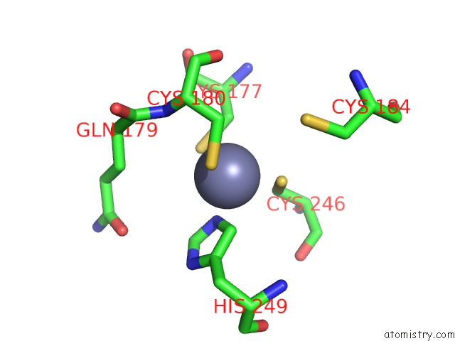
Zinc binding site 1 out
of 6 in 6djx
Go back to
Zinc Binding Sites List in 6djx
Zinc binding site 1 out
of 6 in the Crystal Structure of Pparkin-Pub-UBCH7 Complex
Mono view
Stereo pair view
|
A full contact list of Zinc with other atoms in the Zn binding
site number 1 of Crystal Structure of Pparkin-Pub-UBCH7 Complex within 5.0Å range:
probe
|
atom
|
residue
|
distance (Å)
|
B
|
Occ
|
A:Zn1001
b:0.8
occ:1.00
|
SG
|
A:CYS195
|
2.3
|
0.6
|
1.0
|
SG
|
A:CYS229
|
2.3
|
0.2
|
1.0
|
SG
|
A:CYS222
|
2.3
|
0.6
|
1.0
|
SG
|
A:CYS192
|
2.4
|
0.7
|
1.0
|
CB
|
A:CYS192
|
2.9
|
0.3
|
1.0
|
CB
|
A:CYS222
|
3.0
|
0.3
|
1.0
|
CB
|
A:CYS195
|
3.7
|
0.3
|
1.0
|
CB
|
A:CYS229
|
3.8
|
0.5
|
1.0
|
N
|
A:CYS195
|
4.3
|
0.3
|
1.0
|
CA
|
A:CYS192
|
4.4
|
0.1
|
1.0
|
CA
|
A:CYS222
|
4.5
|
0.1
|
1.0
|
CA
|
A:CYS195
|
4.6
|
0.0
|
1.0
|
O
|
A:GLY197
|
5.0
|
0.7
|
1.0
|
C
|
A:CYS192
|
5.0
|
0.1
|
1.0
|
|
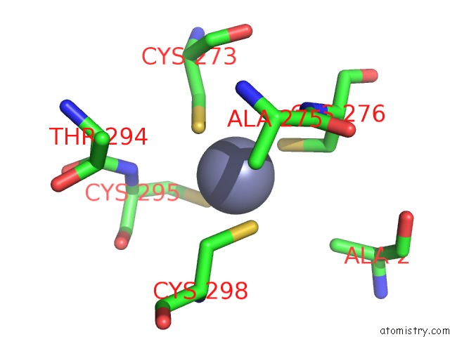
Zinc binding site 2 out
of 6 in 6djx
Go back to
Zinc Binding Sites List in 6djx
Zinc binding site 2 out
of 6 in the Crystal Structure of Pparkin-Pub-UBCH7 Complex
Mono view
Stereo pair view
|
A full contact list of Zinc with other atoms in the Zn binding
site number 2 of Crystal Structure of Pparkin-Pub-UBCH7 Complex within 5.0Å range:
probe
|
atom
|
residue
|
distance (Å)
|
B
|
Occ
|
A:Zn1002
b:0.5
occ:1.00
|
NE2
|
A:HIS249
|
2.0
|
0.4
|
1.0
|
SG
|
A:CYS246
|
2.3
|
0.2
|
1.0
|
SG
|
A:CYS177
|
2.3
|
0.5
|
1.0
|
SG
|
A:CYS180
|
2.3
|
0.9
|
1.0
|
CD2
|
A:HIS249
|
2.4
|
0.0
|
1.0
|
CB
|
A:CYS177
|
2.9
|
0.5
|
1.0
|
CB
|
A:CYS246
|
3.0
|
0.5
|
1.0
|
CE1
|
A:HIS249
|
3.2
|
0.6
|
1.0
|
CB
|
A:CYS180
|
3.3
|
1.0
|
1.0
|
CG
|
A:HIS249
|
3.6
|
0.7
|
1.0
|
N
|
A:CYS180
|
3.9
|
0.2
|
1.0
|
ND1
|
A:HIS249
|
4.0
|
0.8
|
1.0
|
CA
|
A:CYS180
|
4.2
|
0.9
|
1.0
|
CA
|
A:CYS177
|
4.4
|
0.7
|
1.0
|
SG
|
A:CYS184
|
4.4
|
0.3
|
1.0
|
CA
|
A:CYS246
|
4.5
|
0.3
|
1.0
|
CB
|
A:GLN179
|
4.5
|
0.3
|
1.0
|
C
|
A:GLN179
|
4.8
|
0.0
|
1.0
|
CB
|
A:HIS249
|
4.9
|
0.9
|
1.0
|
C
|
A:CYS246
|
4.9
|
0.8
|
1.0
|
N
|
A:GLN179
|
5.0
|
0.9
|
1.0
|
CA
|
A:GLN179
|
5.0
|
0.1
|
1.0
|
|
Zinc binding site 3 out
of 6 in 6djx
Go back to
Zinc Binding Sites List in 6djx
Zinc binding site 3 out
of 6 in the Crystal Structure of Pparkin-Pub-UBCH7 Complex
Mono view
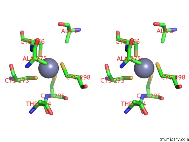
Stereo pair view
|
A full contact list of Zinc with other atoms in the Zn binding
site number 3 of Crystal Structure of Pparkin-Pub-UBCH7 Complex within 5.0Å range:
probe
|
atom
|
residue
|
distance (Å)
|
B
|
Occ
|
A:Zn1003
b:0.1
occ:1.00
|
SG
|
A:CYS298
|
2.3
|
0.5
|
1.0
|
SG
|
A:CYS295
|
2.3
|
0.1
|
1.0
|
SG
|
A:CYS273
|
2.3
|
0.2
|
1.0
|
SG
|
A:CYS276
|
2.3
|
0.6
|
1.0
|
CB
|
A:CYS273
|
3.2
|
1.0
|
1.0
|
CB
|
A:CYS298
|
3.3
|
0.7
|
1.0
|
CB
|
A:CYS276
|
3.5
|
0.3
|
1.0
|
CB
|
A:CYS295
|
3.7
|
0.0
|
1.0
|
N
|
A:CYS276
|
3.8
|
0.0
|
1.0
|
N
|
A:CYS295
|
3.8
|
0.1
|
1.0
|
CB
|
A:ALA275
|
4.1
|
0.4
|
1.0
|
CA
|
A:CYS276
|
4.3
|
0.8
|
1.0
|
CA
|
A:CYS295
|
4.3
|
0.4
|
1.0
|
C
|
A:ALA275
|
4.4
|
0.1
|
1.0
|
N
|
A:CYS298
|
4.4
|
0.9
|
1.0
|
CB
|
C:ALA2
|
4.4
|
0.4
|
1.0
|
CA
|
A:CYS298
|
4.5
|
0.3
|
1.0
|
N
|
A:ALA275
|
4.5
|
0.7
|
1.0
|
CA
|
A:ALA275
|
4.6
|
0.9
|
1.0
|
CA
|
A:CYS273
|
4.6
|
0.7
|
1.0
|
C
|
A:CYS295
|
4.8
|
0.9
|
1.0
|
O
|
A:CYS295
|
4.8
|
0.1
|
1.0
|
C
|
A:CYS273
|
4.9
|
0.3
|
1.0
|
C
|
A:THR294
|
4.9
|
0.4
|
1.0
|
|
Zinc binding site 4 out
of 6 in 6djx
Go back to
Zinc Binding Sites List in 6djx
Zinc binding site 4 out
of 6 in the Crystal Structure of Pparkin-Pub-UBCH7 Complex
Mono view
Stereo pair view
|
A full contact list of Zinc with other atoms in the Zn binding
site number 4 of Crystal Structure of Pparkin-Pub-UBCH7 Complex within 5.0Å range:
probe
|
atom
|
residue
|
distance (Å)
|
B
|
Occ
|
A:Zn1004
b:1.0
occ:1.00
|
ND1
|
A:HIS292
|
2.0
|
0.4
|
1.0
|
SG
|
A:CYS324
|
2.3
|
0.2
|
1.0
|
SG
|
A:CYS328
|
2.3
|
0.4
|
1.0
|
SG
|
A:CYS288
|
2.3
|
0.9
|
1.0
|
CE1
|
A:HIS292
|
2.6
|
0.5
|
1.0
|
CB
|
A:CYS328
|
3.0
|
0.6
|
1.0
|
CB
|
A:CYS288
|
3.2
|
0.1
|
1.0
|
CG
|
A:HIS292
|
3.2
|
0.9
|
1.0
|
CB
|
A:CYS324
|
3.4
|
0.6
|
1.0
|
NE2
|
A:HIS292
|
3.8
|
0.6
|
1.0
|
CB
|
A:HIS292
|
3.9
|
0.4
|
1.0
|
CD2
|
A:HIS292
|
4.1
|
0.1
|
1.0
|
CA
|
A:CYS328
|
4.2
|
0.2
|
1.0
|
N
|
A:CYS328
|
4.3
|
0.1
|
1.0
|
CB
|
A:ALA326
|
4.4
|
0.1
|
1.0
|
CA
|
A:CYS288
|
4.6
|
0.2
|
1.0
|
N
|
A:HIS292
|
4.8
|
0.9
|
1.0
|
CB
|
A:SER290
|
4.8
|
0.7
|
1.0
|
CA
|
A:CYS324
|
4.9
|
0.1
|
1.0
|
OG
|
A:SER331
|
4.9
|
0.2
|
1.0
|
CA
|
A:HIS292
|
5.0
|
0.9
|
1.0
|
|
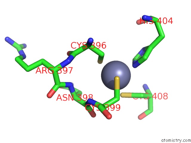
Zinc binding site 5 out
of 6 in 6djx
Go back to
Zinc Binding Sites List in 6djx
Zinc binding site 5 out
of 6 in the Crystal Structure of Pparkin-Pub-UBCH7 Complex
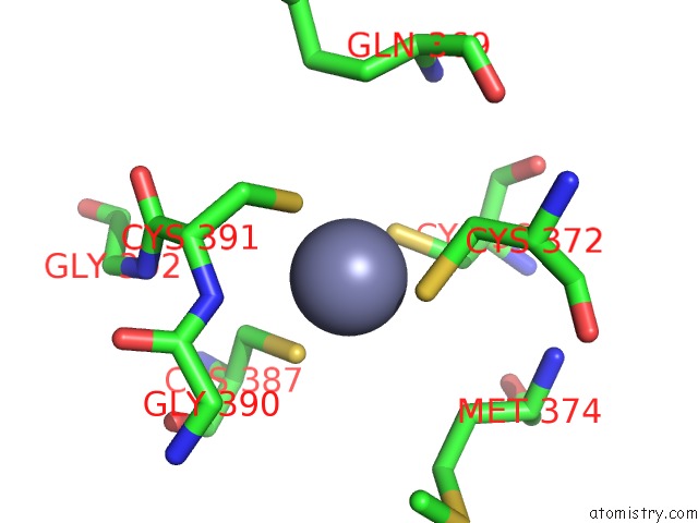
Mono view
Stereo pair view
|
A full contact list of Zinc with other atoms in the Zn binding
site number 5 of Crystal Structure of Pparkin-Pub-UBCH7 Complex within 5.0Å range:
probe
|
atom
|
residue
|
distance (Å)
|
B
|
Occ
|
A:Zn1005
b:0.1
occ:1.00
|
NE2
|
A:HIS404
|
2.0
|
0.6
|
1.0
|
SG
|
A:CYS396
|
2.2
|
0.9
|
1.0
|
SG
|
A:CYS399
|
2.3
|
0.3
|
1.0
|
SG
|
A:CYS408
|
2.3
|
0.9
|
1.0
|
CB
|
A:CYS396
|
2.5
|
0.2
|
1.0
|
CE1
|
A:HIS404
|
2.6
|
0.9
|
1.0
|
CB
|
A:CYS399
|
2.8
|
0.2
|
1.0
|
CB
|
A:CYS408
|
3.2
|
0.3
|
1.0
|
CD2
|
A:HIS404
|
3.2
|
0.7
|
1.0
|
N
|
A:CYS399
|
3.6
|
0.7
|
1.0
|
CA
|
A:CYS399
|
3.8
|
0.9
|
1.0
|
ND1
|
A:HIS404
|
3.8
|
0.7
|
1.0
|
C
|
A:ASN398
|
3.9
|
0.2
|
1.0
|
CA
|
A:CYS396
|
3.9
|
0.4
|
1.0
|
CG
|
A:HIS404
|
4.1
|
0.0
|
1.0
|
O
|
A:ASN398
|
4.3
|
1.0
|
1.0
|
CB
|
A:ASN398
|
4.3
|
0.8
|
1.0
|
N
|
A:ASN398
|
4.4
|
0.7
|
1.0
|
N
|
A:CYS408
|
4.4
|
0.7
|
1.0
|
CA
|
A:CYS408
|
4.4
|
0.8
|
1.0
|
CA
|
A:ASN398
|
4.4
|
0.3
|
1.0
|
C
|
A:CYS396
|
4.4
|
0.1
|
1.0
|
N
|
A:CYS396
|
4.7
|
0.2
|
1.0
|
O
|
A:CYS396
|
4.8
|
0.5
|
1.0
|
C
|
A:CYS399
|
4.8
|
0.5
|
1.0
|
N
|
A:ARG397
|
4.9
|
0.8
|
1.0
|
|
Zinc binding site 6 out
of 6 in 6djx
Go back to
Zinc Binding Sites List in 6djx
Zinc binding site 6 out
of 6 in the Crystal Structure of Pparkin-Pub-UBCH7 Complex
Mono view
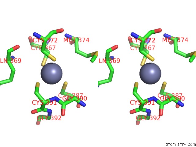
Stereo pair view
|
A full contact list of Zinc with other atoms in the Zn binding
site number 6 of Crystal Structure of Pparkin-Pub-UBCH7 Complex within 5.0Å range:
probe
|
atom
|
residue
|
distance (Å)
|
B
|
Occ
|
A:Zn1006
b:0.5
occ:1.00
|
SG
|
A:CYS391
|
2.2
|
0.1
|
1.0
|
SG
|
A:CYS367
|
2.3
|
0.8
|
1.0
|
SG
|
A:CYS372
|
2.3
|
1.0
|
1.0
|
SG
|
A:CYS387
|
2.3
|
0.1
|
1.0
|
CB
|
A:CYS367
|
3.4
|
0.3
|
1.0
|
CB
|
A:CYS387
|
3.4
|
0.1
|
1.0
|
CB
|
A:CYS391
|
3.5
|
0.2
|
1.0
|
CB
|
A:CYS372
|
3.5
|
0.2
|
1.0
|
N
|
A:CYS391
|
3.5
|
0.3
|
1.0
|
CA
|
A:CYS391
|
4.0
|
0.9
|
1.0
|
CB
|
A:MET374
|
4.4
|
0.5
|
1.0
|
C
|
A:GLY390
|
4.6
|
0.3
|
1.0
|
C
|
A:CYS391
|
4.7
|
0.4
|
1.0
|
CA
|
A:GLY390
|
4.7
|
1.0
|
1.0
|
CA
|
A:CYS387
|
4.8
|
0.3
|
1.0
|
CA
|
A:CYS367
|
4.8
|
0.3
|
1.0
|
N
|
A:GLY392
|
4.8
|
0.1
|
1.0
|
CB
|
A:GLN369
|
4.9
|
0.2
|
1.0
|
CA
|
A:CYS372
|
4.9
|
0.7
|
1.0
|
|
Reference:
V.Sauve,
G.Sung,
N.Soya,
G.Kozlov,
N.Blaimschein,
L.S.Miotto,
J.F.Trempe,
G.L.Lukacs,
K.Gehring.
Mechanism of Parkin Activation By Phosphorylation. Nat. Struct. Mol. Biol. V. 25 623 2018.
ISSN: ESSN 1545-9985
PubMed: 29967542
DOI: 10.1038/S41594-018-0088-7
Page generated: Mon Oct 28 19:35:35 2024
|