Zinc »
PDB 6d5s-6diu »
6ddr »
Zinc in PDB 6ddr: Crystal Structure Analysis of the Epitope of An Anti-Mica Antibody
Protein crystallography data
The structure of Crystal Structure Analysis of the Epitope of An Anti-Mica Antibody, PDB code: 6ddr
was solved by
M.L.Matsumoto,
with X-Ray Crystallography technique. A brief refinement statistics is given in the table below:
| Resolution Low / High (Å) | 35.00 / 1.90 |
| Space group | P 21 21 21 |
| Cell size a, b, c (Å), α, β, γ (°) | 51.549, 61.833, 172.368, 90.00, 90.00, 90.00 |
| R / Rfree (%) | 20.8 / 24.7 |
Zinc Binding Sites:
The binding sites of Zinc atom in the Crystal Structure Analysis of the Epitope of An Anti-Mica Antibody
(pdb code 6ddr). This binding sites where shown within
5.0 Angstroms radius around Zinc atom.
In total only one binding site of Zinc was determined in the Crystal Structure Analysis of the Epitope of An Anti-Mica Antibody, PDB code: 6ddr:
In total only one binding site of Zinc was determined in the Crystal Structure Analysis of the Epitope of An Anti-Mica Antibody, PDB code: 6ddr:
Zinc binding site 1 out of 1 in 6ddr
Go back to
Zinc binding site 1 out
of 1 in the Crystal Structure Analysis of the Epitope of An Anti-Mica Antibody
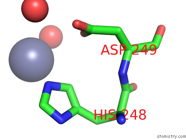
Mono view
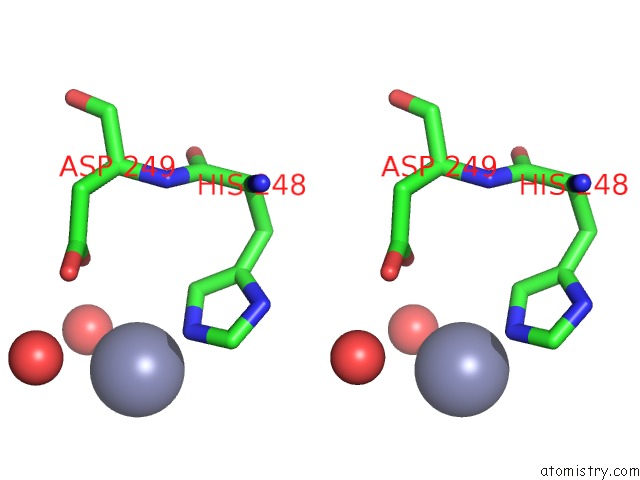
Stereo pair view
Mono view
Stereo pair view
A full contact list of Zinc with other atoms in the Zn binding
site number 1 of Crystal Structure Analysis of the Epitope of An Anti-Mica Antibody within 5.0Å range:
|
Reference:
T.N.Lombana,
M.L.Matsumoto,
A.M.Berkley,
E.Toy,
R.Cook,
Y.Gan,
C.Du,
P.Schnier,
W.Sandoval,
Z.Ye,
J.M.Schartner,
J.Kim,
C.Spiess.
High-Resolution Glycosylation Site-Engineering Method Identifies Mica Epitope Critical For Shedding Inhibition Activity of Anti-Mica Antibodies. Mabs V. 11 75 2019.
ISSN: ESSN 1942-0870
PubMed: 30307368
DOI: 10.1080/19420862.2018.1532767
Page generated: Mon Oct 28 19:27:45 2024
ISSN: ESSN 1942-0870
PubMed: 30307368
DOI: 10.1080/19420862.2018.1532767
Last articles
Zn in 9MJ5Zn in 9HNW
Zn in 9G0L
Zn in 9FNE
Zn in 9DZN
Zn in 9E0I
Zn in 9D32
Zn in 9DAK
Zn in 8ZXC
Zn in 8ZUF