Zinc »
PDB 6cln-6cvt »
6cty »
Zinc in PDB 6cty: Crystal Structure of Dihydroorotase Pyrc From Yersinia Pestis in Complex with Zinc and Malate at 2.4 A Resolution
Enzymatic activity of Crystal Structure of Dihydroorotase Pyrc From Yersinia Pestis in Complex with Zinc and Malate at 2.4 A Resolution
All present enzymatic activity of Crystal Structure of Dihydroorotase Pyrc From Yersinia Pestis in Complex with Zinc and Malate at 2.4 A Resolution:
3.5.2.3;
3.5.2.3;
Protein crystallography data
The structure of Crystal Structure of Dihydroorotase Pyrc From Yersinia Pestis in Complex with Zinc and Malate at 2.4 A Resolution, PDB code: 6cty
was solved by
J.Lipowska,
I.G.Shabalin,
J.Winsor,
M.Woinska,
D.R.Cooper,
K.Kwon,
L.Shuvalova,
W.F.Anderson,
W.Minor,
Center For Structural Genomics Ofinfectious Diseases (Csgid),
with X-Ray Crystallography technique. A brief refinement statistics is given in the table below:
| Resolution Low / High (Å) | 50.01 / 2.41 |
| Space group | P 21 21 21 |
| Cell size a, b, c (Å), α, β, γ (°) | 95.838, 112.202, 208.264, 90.00, 90.00, 90.00 |
| R / Rfree (%) | 15.9 / 19.8 |
Zinc Binding Sites:
Pages:
>>> Page 1 <<< Page 2, Binding sites: 11 - 12;Binding sites:
The binding sites of Zinc atom in the Crystal Structure of Dihydroorotase Pyrc From Yersinia Pestis in Complex with Zinc and Malate at 2.4 A Resolution (pdb code 6cty). This binding sites where shown within 5.0 Angstroms radius around Zinc atom.In total 12 binding sites of Zinc where determined in the Crystal Structure of Dihydroorotase Pyrc From Yersinia Pestis in Complex with Zinc and Malate at 2.4 A Resolution, PDB code: 6cty:
Jump to Zinc binding site number: 1; 2; 3; 4; 5; 6; 7; 8; 9; 10;
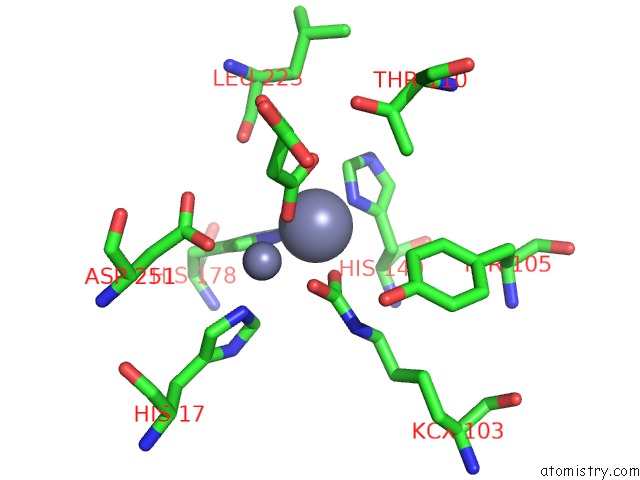
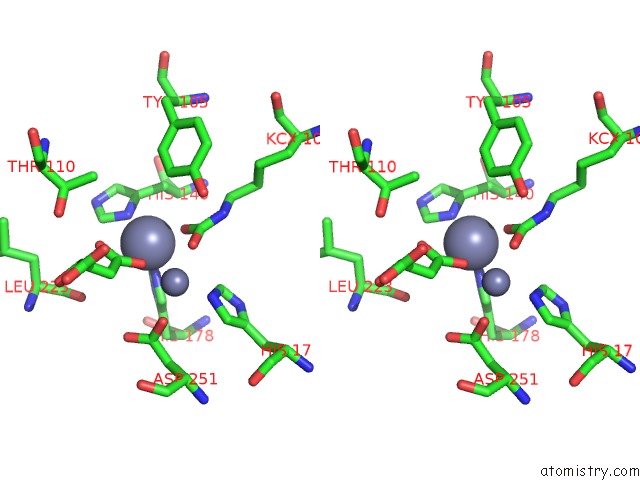
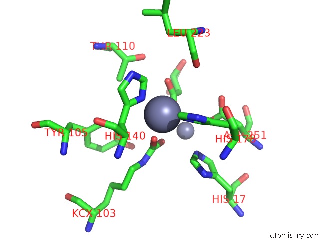
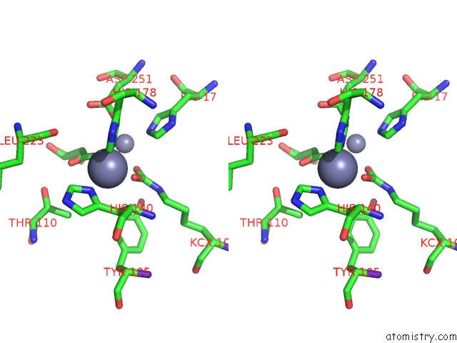
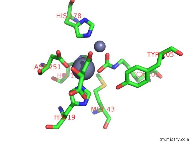
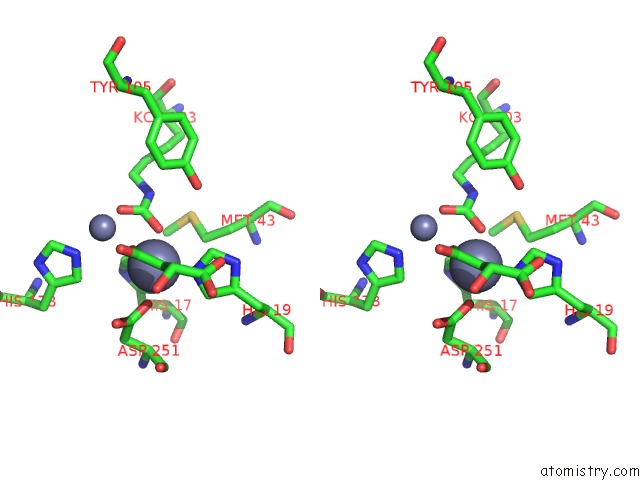
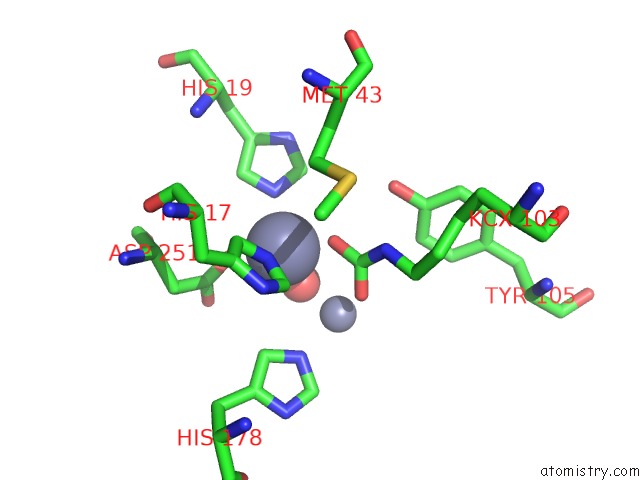
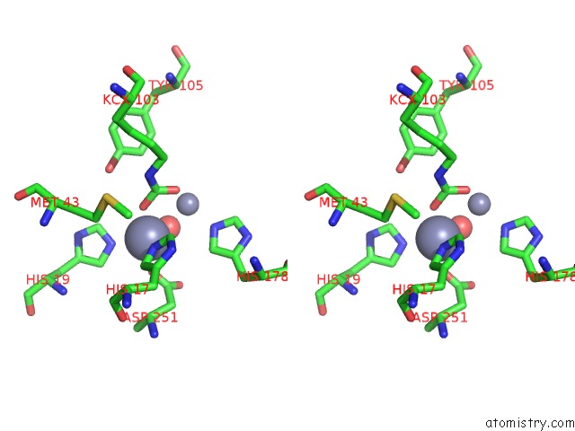
Zinc binding site 1 out of 12 in 6cty
Go back to
Zinc binding site 1 out
of 12 in the Crystal Structure of Dihydroorotase Pyrc From Yersinia Pestis in Complex with Zinc and Malate at 2.4 A Resolution
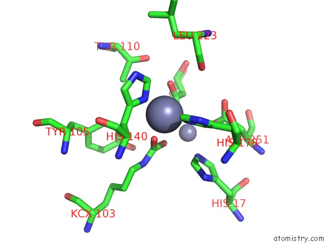
Mono view
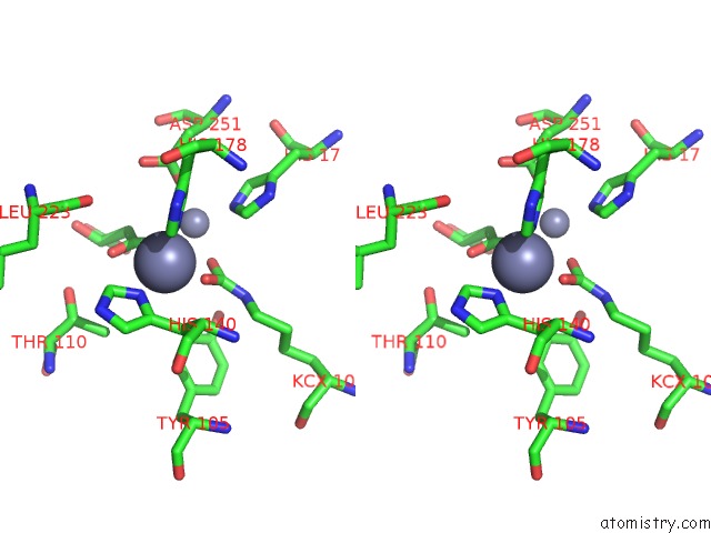
Stereo pair view
Mono view
Stereo pair view
A full contact list of Zinc with other atoms in the Zn binding
site number 1 of Crystal Structure of Dihydroorotase Pyrc From Yersinia Pestis in Complex with Zinc and Malate at 2.4 A Resolution within 5.0Å range:
|
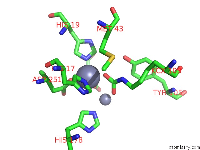
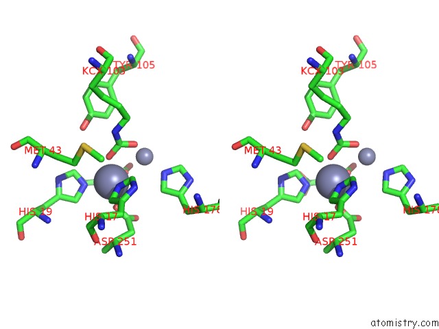
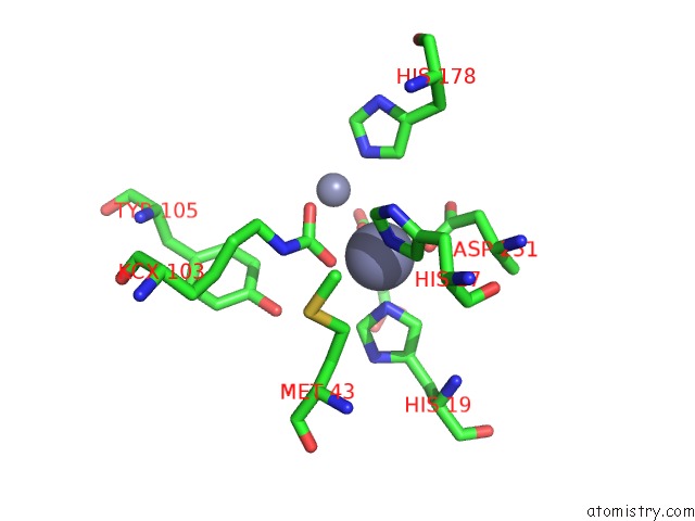
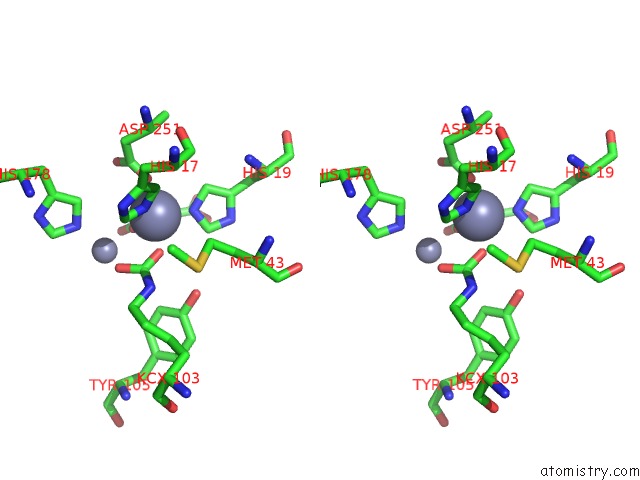
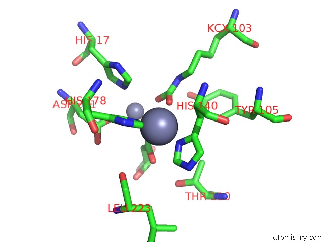
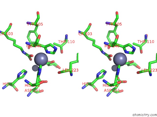
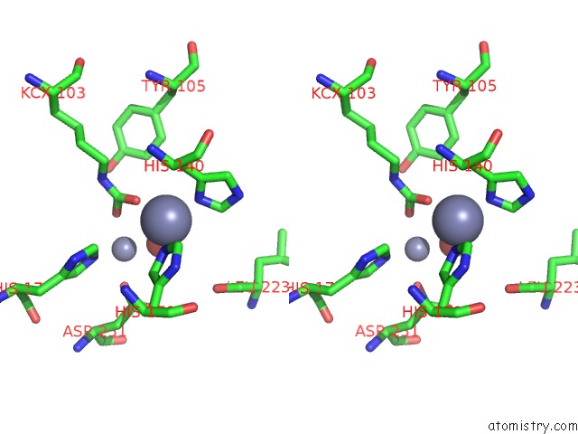
Zinc binding site 2 out of 12 in 6cty
Go back to
Zinc binding site 2 out
of 12 in the Crystal Structure of Dihydroorotase Pyrc From Yersinia Pestis in Complex with Zinc and Malate at 2.4 A Resolution
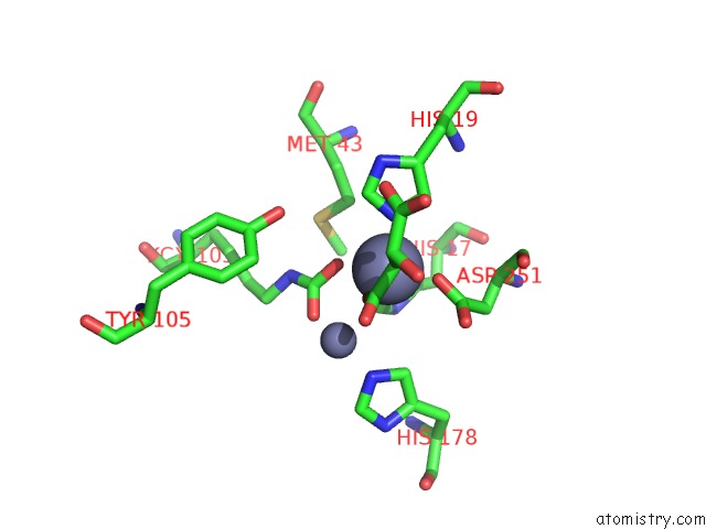
Mono view
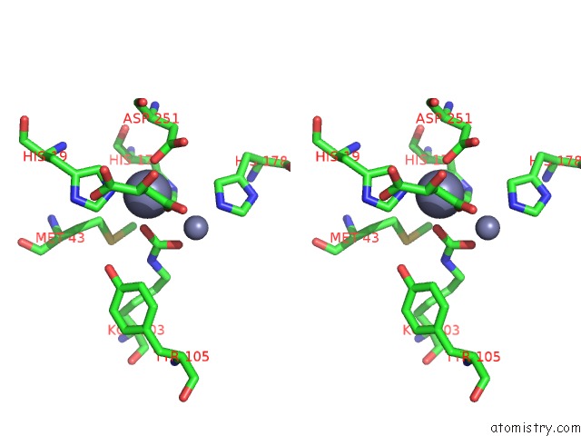
Stereo pair view
Mono view
Stereo pair view
A full contact list of Zinc with other atoms in the Zn binding
site number 2 of Crystal Structure of Dihydroorotase Pyrc From Yersinia Pestis in Complex with Zinc and Malate at 2.4 A Resolution within 5.0Å range:
|
Zinc binding site 3 out of 12 in 6cty
Go back to
Zinc binding site 3 out
of 12 in the Crystal Structure of Dihydroorotase Pyrc From Yersinia Pestis in Complex with Zinc and Malate at 2.4 A Resolution
Mono view
Stereo pair view
Mono view
Stereo pair view
A full contact list of Zinc with other atoms in the Zn binding
site number 3 of Crystal Structure of Dihydroorotase Pyrc From Yersinia Pestis in Complex with Zinc and Malate at 2.4 A Resolution within 5.0Å range:
|
Zinc binding site 4 out of 12 in 6cty
Go back to
Zinc binding site 4 out
of 12 in the Crystal Structure of Dihydroorotase Pyrc From Yersinia Pestis in Complex with Zinc and Malate at 2.4 A Resolution
Mono view
Stereo pair view
Mono view
Stereo pair view
A full contact list of Zinc with other atoms in the Zn binding
site number 4 of Crystal Structure of Dihydroorotase Pyrc From Yersinia Pestis in Complex with Zinc and Malate at 2.4 A Resolution within 5.0Å range:
|
Zinc binding site 5 out of 12 in 6cty
Go back to
Zinc binding site 5 out
of 12 in the Crystal Structure of Dihydroorotase Pyrc From Yersinia Pestis in Complex with Zinc and Malate at 2.4 A Resolution
Mono view
Stereo pair view
Mono view
Stereo pair view
A full contact list of Zinc with other atoms in the Zn binding
site number 5 of Crystal Structure of Dihydroorotase Pyrc From Yersinia Pestis in Complex with Zinc and Malate at 2.4 A Resolution within 5.0Å range:
|
Zinc binding site 6 out of 12 in 6cty
Go back to
Zinc binding site 6 out
of 12 in the Crystal Structure of Dihydroorotase Pyrc From Yersinia Pestis in Complex with Zinc and Malate at 2.4 A Resolution
Mono view
Stereo pair view
Mono view
Stereo pair view
A full contact list of Zinc with other atoms in the Zn binding
site number 6 of Crystal Structure of Dihydroorotase Pyrc From Yersinia Pestis in Complex with Zinc and Malate at 2.4 A Resolution within 5.0Å range:
|
Zinc binding site 7 out of 12 in 6cty
Go back to
Zinc binding site 7 out
of 12 in the Crystal Structure of Dihydroorotase Pyrc From Yersinia Pestis in Complex with Zinc and Malate at 2.4 A Resolution
Mono view
Stereo pair view
Mono view
Stereo pair view
A full contact list of Zinc with other atoms in the Zn binding
site number 7 of Crystal Structure of Dihydroorotase Pyrc From Yersinia Pestis in Complex with Zinc and Malate at 2.4 A Resolution within 5.0Å range:
|
Zinc binding site 8 out of 12 in 6cty
Go back to
Zinc binding site 8 out
of 12 in the Crystal Structure of Dihydroorotase Pyrc From Yersinia Pestis in Complex with Zinc and Malate at 2.4 A Resolution
Mono view
Stereo pair view
Mono view
Stereo pair view
A full contact list of Zinc with other atoms in the Zn binding
site number 8 of Crystal Structure of Dihydroorotase Pyrc From Yersinia Pestis in Complex with Zinc and Malate at 2.4 A Resolution within 5.0Å range:
|
Zinc binding site 9 out of 12 in 6cty
Go back to
Zinc binding site 9 out
of 12 in the Crystal Structure of Dihydroorotase Pyrc From Yersinia Pestis in Complex with Zinc and Malate at 2.4 A Resolution
Mono view
Stereo pair view
Mono view
Stereo pair view
A full contact list of Zinc with other atoms in the Zn binding
site number 9 of Crystal Structure of Dihydroorotase Pyrc From Yersinia Pestis in Complex with Zinc and Malate at 2.4 A Resolution within 5.0Å range:
|
Zinc binding site 10 out of 12 in 6cty
Go back to
Zinc binding site 10 out
of 12 in the Crystal Structure of Dihydroorotase Pyrc From Yersinia Pestis in Complex with Zinc and Malate at 2.4 A Resolution

Mono view
Stereo pair view
Mono view
Stereo pair view
A full contact list of Zinc with other atoms in the Zn binding
site number 10 of Crystal Structure of Dihydroorotase Pyrc From Yersinia Pestis in Complex with Zinc and Malate at 2.4 A Resolution within 5.0Å range:
|
Reference:
J.Lipowska,
C.D.Miks,
K.Kwon,
L.Shuvalova,
H.Zheng,
K.Lewinski,
D.R.Cooper,
I.G.Shabalin,
W.Minor.
Pyrimidine Biosynthesis in Pathogens - Structures and Analysis of Dihydroorotases From Yersinia Pestis and Vibrio Cholerae. Int.J.Biol.Macromol. V. 136 1176 2019.
ISSN: ISSN 0141-8130
PubMed: 31207330
DOI: 10.1016/J.IJBIOMAC.2019.05.149
Page generated: Mon Oct 28 19:09:50 2024
ISSN: ISSN 0141-8130
PubMed: 31207330
DOI: 10.1016/J.IJBIOMAC.2019.05.149
Last articles
Zn in 9MJ5Zn in 9HNW
Zn in 9G0L
Zn in 9FNE
Zn in 9DZN
Zn in 9E0I
Zn in 9D32
Zn in 9DAK
Zn in 8ZXC
Zn in 8ZUF