Zinc »
PDB 6bh7-6bsm »
6bn7 »
Zinc in PDB 6bn7: Crystal Structure of DDB1-Crbn-BRD4(BD1) Complex Bound to DBET23 Protac.
Protein crystallography data
The structure of Crystal Structure of DDB1-Crbn-BRD4(BD1) Complex Bound to DBET23 Protac., PDB code: 6bn7
was solved by
R.P.Nowak,
S.L.Deangelo,
D.Buckley,
J.E.Bradner,
E.S.Fischer,
with X-Ray Crystallography technique. A brief refinement statistics is given in the table below:
| Resolution Low / High (Å) | 49.87 / 3.50 |
| Space group | P 65 2 2 |
| Cell size a, b, c (Å), α, β, γ (°) | 115.571, 115.571, 596.315, 90.00, 90.00, 120.00 |
| R / Rfree (%) | 21.2 / 25.6 |
Other elements in 6bn7:
The structure of Crystal Structure of DDB1-Crbn-BRD4(BD1) Complex Bound to DBET23 Protac. also contains other interesting chemical elements:
| Chlorine | (Cl) | 1 atom |
Zinc Binding Sites:
The binding sites of Zinc atom in the Crystal Structure of DDB1-Crbn-BRD4(BD1) Complex Bound to DBET23 Protac.
(pdb code 6bn7). This binding sites where shown within
5.0 Angstroms radius around Zinc atom.
In total only one binding site of Zinc was determined in the Crystal Structure of DDB1-Crbn-BRD4(BD1) Complex Bound to DBET23 Protac., PDB code: 6bn7:
In total only one binding site of Zinc was determined in the Crystal Structure of DDB1-Crbn-BRD4(BD1) Complex Bound to DBET23 Protac., PDB code: 6bn7:
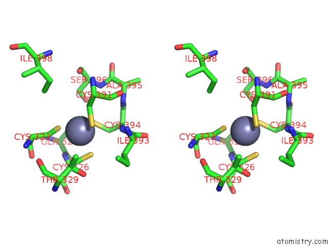
Zinc binding site 1 out of 1 in 6bn7
Go back to
Zinc binding site 1 out
of 1 in the Crystal Structure of DDB1-Crbn-BRD4(BD1) Complex Bound to DBET23 Protac.
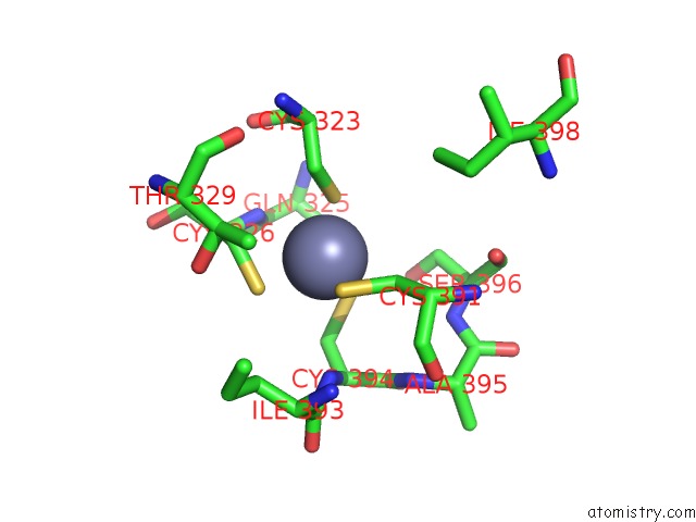
Mono view
Stereo pair view
Mono view
Stereo pair view
A full contact list of Zinc with other atoms in the Zn binding
site number 1 of Crystal Structure of DDB1-Crbn-BRD4(BD1) Complex Bound to DBET23 Protac. within 5.0Å range:
|
Reference:
R.P.Nowak,
S.L.Deangelo,
D.Buckley,
Z.He,
K.A.Donovan,
J.An,
N.Safaee,
M.P.Jedrychowski,
C.M.Ponthier,
M.Ishoey,
T.Zhang,
J.D.Mancias,
N.S.Gray,
J.E.Bradner,
E.S.Fischer.
Plasticity in Binding Confers Selectivity in Ligand-Induced Protein Degradation. Nat. Chem. Biol. V. 14 706 2018.
ISSN: ESSN 1552-4469
PubMed: 29892083
DOI: 10.1038/S41589-018-0055-Y
Page generated: Mon Oct 28 18:08:32 2024
ISSN: ESSN 1552-4469
PubMed: 29892083
DOI: 10.1038/S41589-018-0055-Y
Last articles
Zn in 9MJ5Zn in 9HNW
Zn in 9G0L
Zn in 9FNE
Zn in 9DZN
Zn in 9E0I
Zn in 9D32
Zn in 9DAK
Zn in 8ZXC
Zn in 8ZUF