Zinc »
PDB 6av3-6be6 »
6b05 »
Zinc in PDB 6b05: The Crystal Structure of the Ferredoxin Protease Fusc E83A Mutant in Complex with Arabidopsis Ferredoxin
Protein crystallography data
The structure of The Crystal Structure of the Ferredoxin Protease Fusc E83A Mutant in Complex with Arabidopsis Ferredoxin, PDB code: 6b05
was solved by
R.Grinter,
with X-Ray Crystallography technique. A brief refinement statistics is given in the table below:
| Resolution Low / High (Å) | 39.99 / 1.90 |
| Space group | P 2 21 21 |
| Cell size a, b, c (Å), α, β, γ (°) | 81.338, 126.505, 133.545, 90.00, 90.00, 90.00 |
| R / Rfree (%) | 19.5 / 23.9 |
Zinc Binding Sites:
The binding sites of Zinc atom in the The Crystal Structure of the Ferredoxin Protease Fusc E83A Mutant in Complex with Arabidopsis Ferredoxin
(pdb code 6b05). This binding sites where shown within
5.0 Angstroms radius around Zinc atom.
In total only one binding site of Zinc was determined in the The Crystal Structure of the Ferredoxin Protease Fusc E83A Mutant in Complex with Arabidopsis Ferredoxin, PDB code: 6b05:
In total only one binding site of Zinc was determined in the The Crystal Structure of the Ferredoxin Protease Fusc E83A Mutant in Complex with Arabidopsis Ferredoxin, PDB code: 6b05:
Zinc binding site 1 out of 1 in 6b05
Go back to
Zinc binding site 1 out
of 1 in the The Crystal Structure of the Ferredoxin Protease Fusc E83A Mutant in Complex with Arabidopsis Ferredoxin

Mono view
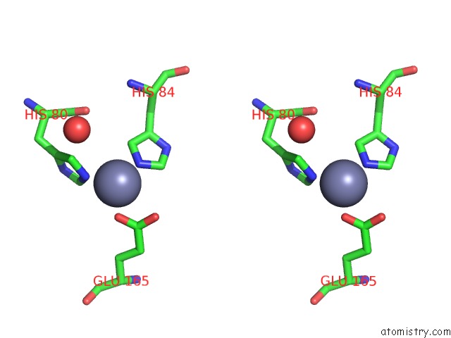
Stereo pair view
Mono view
Stereo pair view
A full contact list of Zinc with other atoms in the Zn binding
site number 1 of The Crystal Structure of the Ferredoxin Protease Fusc E83A Mutant in Complex with Arabidopsis Ferredoxin within 5.0Å range:
|
Reference:
R.Grinter,
I.D.Hay,
J.Song,
J.Wang,
D.Teng,
V.Dhanesakaran,
J.J.Wilksch,
M.R.Davies,
D.Littler,
S.A.Beckham,
I.R.Henderson,
R.A.Strugnell,
G.Dougan,
T.Lithgow.
Fusc, A Member of the M16 Protease Family Acquired By Bacteria For Iron Piracy Against Plants. Plos Biol. V. 16 06026 2018.
ISSN: ESSN 1545-7885
PubMed: 30071011
DOI: 10.1371/JOURNAL.PBIO.2006026
Page generated: Mon Oct 28 17:45:51 2024
ISSN: ESSN 1545-7885
PubMed: 30071011
DOI: 10.1371/JOURNAL.PBIO.2006026
Last articles
Zn in 9MJ5Zn in 9HNW
Zn in 9G0L
Zn in 9FNE
Zn in 9DZN
Zn in 9E0I
Zn in 9D32
Zn in 9DAK
Zn in 8ZXC
Zn in 8ZUF