Zinc »
PDB 6a57-6agg »
6aem »
Zinc in PDB 6aem: Crystal Structure of the PKD1 Domain of Vibrio Anguillarum Epp
Protein crystallography data
The structure of Crystal Structure of the PKD1 Domain of Vibrio Anguillarum Epp, PDB code: 6aem
was solved by
Q.Ma,
P.Li,
with X-Ray Crystallography technique. A brief refinement statistics is given in the table below:
| Resolution Low / High (Å) | 35.25 / 1.27 |
| Space group | C 1 2 1 |
| Cell size a, b, c (Å), α, β, γ (°) | 71.096, 46.069, 41.616, 90.00, 97.39, 90.00 |
| R / Rfree (%) | 15.1 / 18.6 |
Other elements in 6aem:
The structure of Crystal Structure of the PKD1 Domain of Vibrio Anguillarum Epp also contains other interesting chemical elements:
| Calcium | (Ca) | 2 atoms |
Zinc Binding Sites:
The binding sites of Zinc atom in the Crystal Structure of the PKD1 Domain of Vibrio Anguillarum Epp
(pdb code 6aem). This binding sites where shown within
5.0 Angstroms radius around Zinc atom.
In total 5 binding sites of Zinc where determined in the Crystal Structure of the PKD1 Domain of Vibrio Anguillarum Epp, PDB code: 6aem:
Jump to Zinc binding site number: 1; 2; 3; 4; 5;
In total 5 binding sites of Zinc where determined in the Crystal Structure of the PKD1 Domain of Vibrio Anguillarum Epp, PDB code: 6aem:
Jump to Zinc binding site number: 1; 2; 3; 4; 5;
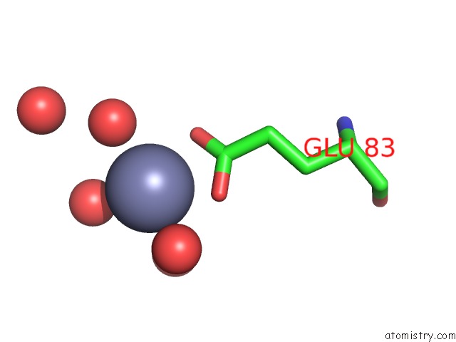
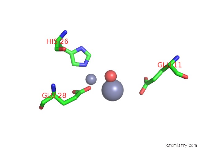
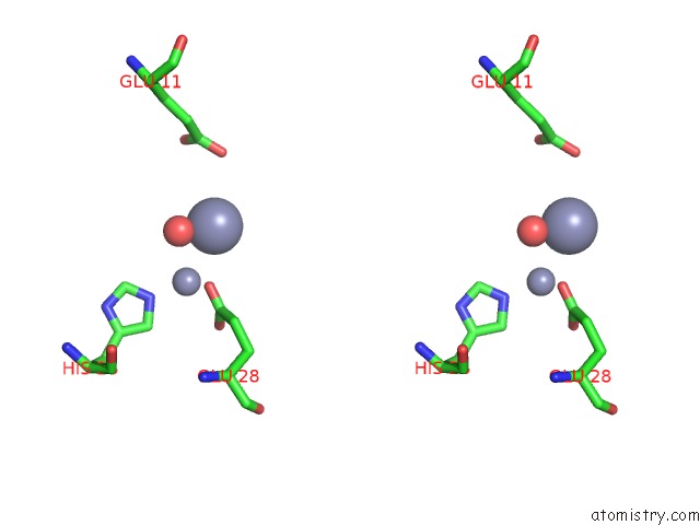
Zinc binding site 1 out of 5 in 6aem
Go back to
Zinc binding site 1 out
of 5 in the Crystal Structure of the PKD1 Domain of Vibrio Anguillarum Epp
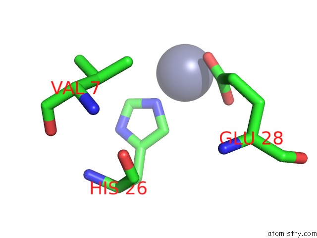
Mono view
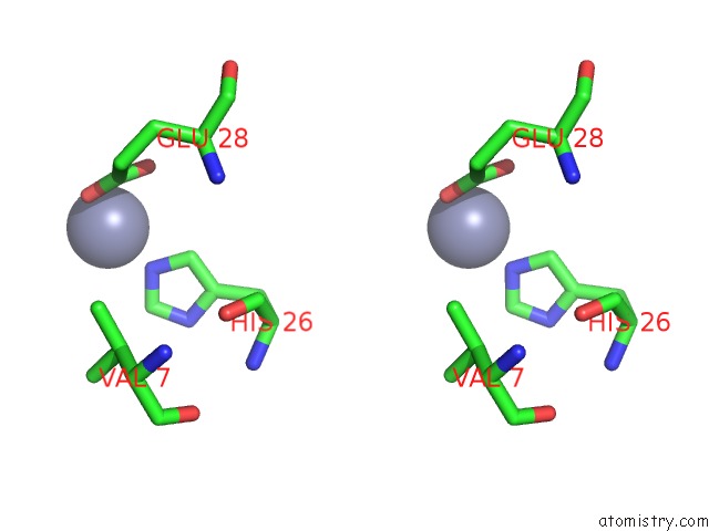
Stereo pair view
Mono view
Stereo pair view
A full contact list of Zinc with other atoms in the Zn binding
site number 1 of Crystal Structure of the PKD1 Domain of Vibrio Anguillarum Epp within 5.0Å range:
|
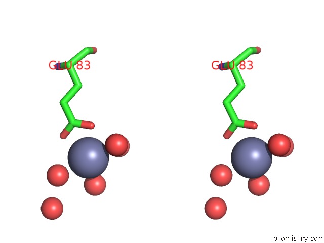
Zinc binding site 2 out of 5 in 6aem
Go back to
Zinc binding site 2 out
of 5 in the Crystal Structure of the PKD1 Domain of Vibrio Anguillarum Epp
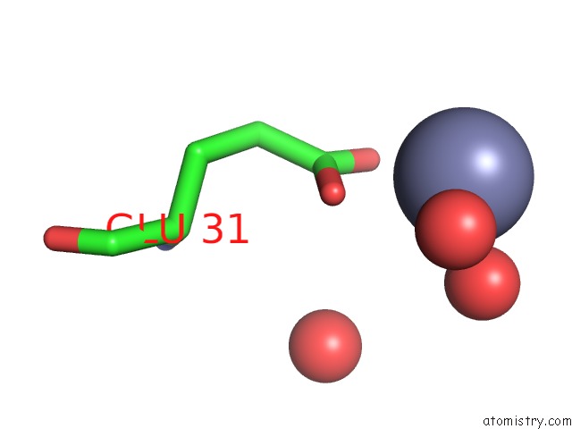
Mono view
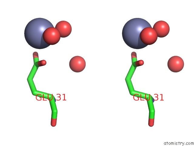
Stereo pair view
Mono view
Stereo pair view
A full contact list of Zinc with other atoms in the Zn binding
site number 2 of Crystal Structure of the PKD1 Domain of Vibrio Anguillarum Epp within 5.0Å range:
|
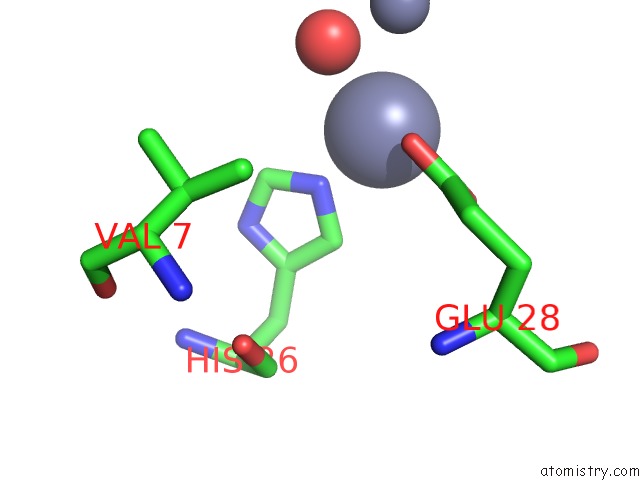
Zinc binding site 3 out of 5 in 6aem
Go back to
Zinc binding site 3 out
of 5 in the Crystal Structure of the PKD1 Domain of Vibrio Anguillarum Epp
Mono view
Stereo pair view
Mono view
Stereo pair view
A full contact list of Zinc with other atoms in the Zn binding
site number 3 of Crystal Structure of the PKD1 Domain of Vibrio Anguillarum Epp within 5.0Å range:
|
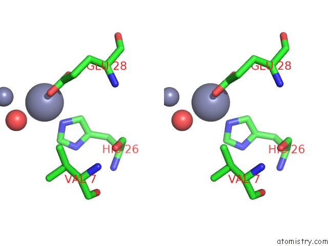
Zinc binding site 4 out of 5 in 6aem
Go back to
Zinc binding site 4 out
of 5 in the Crystal Structure of the PKD1 Domain of Vibrio Anguillarum Epp
Mono view
Stereo pair view
Mono view
Stereo pair view
A full contact list of Zinc with other atoms in the Zn binding
site number 4 of Crystal Structure of the PKD1 Domain of Vibrio Anguillarum Epp within 5.0Å range:
|
Zinc binding site 5 out of 5 in 6aem
Go back to
Zinc binding site 5 out
of 5 in the Crystal Structure of the PKD1 Domain of Vibrio Anguillarum Epp
Mono view
Stereo pair view
Mono view
Stereo pair view
A full contact list of Zinc with other atoms in the Zn binding
site number 5 of Crystal Structure of the PKD1 Domain of Vibrio Anguillarum Epp within 5.0Å range:
|
Reference:
P.Li,
K.Zang,
Y.Li,
C.Liu,
Q.Ma.
Structural Basis For Specific Calcium Binding By the Polycystic-Kidney-Disease Domain of Vibrio Anguillarum Protease Epp Biochem. Biophys. Res. V. 505 471 2018COMMUN..
ISSN: ESSN 1090-2104
PubMed: 30268503
DOI: 10.1016/J.BBRC.2018.09.108
Page generated: Mon Oct 28 17:29:30 2024
ISSN: ESSN 1090-2104
PubMed: 30268503
DOI: 10.1016/J.BBRC.2018.09.108
Last articles
Zn in 9MJ5Zn in 9HNW
Zn in 9G0L
Zn in 9FNE
Zn in 9DZN
Zn in 9E0I
Zn in 9D32
Zn in 9DAK
Zn in 8ZXC
Zn in 8ZUF