Zinc »
PDB 5yo1-5yy0 »
5yxc »
Zinc in PDB 5yxc: Crystal Structure of Zinc Binding Protein Zint in Complex with Citrate From E. Coli
Protein crystallography data
The structure of Crystal Structure of Zinc Binding Protein Zint in Complex with Citrate From E. Coli, PDB code: 5yxc
was solved by
J.Chen,
L.Wang,
F.Shang,
Y.Xu,
with X-Ray Crystallography technique. A brief refinement statistics is given in the table below:
| Resolution Low / High (Å) | 41.03 / 1.76 |
| Space group | P 1 21 1 |
| Cell size a, b, c (Å), α, β, γ (°) | 41.040, 65.866, 73.536, 90.00, 91.35, 90.00 |
| R / Rfree (%) | 18.8 / 23.4 |
Zinc Binding Sites:
The binding sites of Zinc atom in the Crystal Structure of Zinc Binding Protein Zint in Complex with Citrate From E. Coli
(pdb code 5yxc). This binding sites where shown within
5.0 Angstroms radius around Zinc atom.
In total 2 binding sites of Zinc where determined in the Crystal Structure of Zinc Binding Protein Zint in Complex with Citrate From E. Coli, PDB code: 5yxc:
Jump to Zinc binding site number: 1; 2;
In total 2 binding sites of Zinc where determined in the Crystal Structure of Zinc Binding Protein Zint in Complex with Citrate From E. Coli, PDB code: 5yxc:
Jump to Zinc binding site number: 1; 2;
Zinc binding site 1 out of 2 in 5yxc
Go back to
Zinc binding site 1 out
of 2 in the Crystal Structure of Zinc Binding Protein Zint in Complex with Citrate From E. Coli

Mono view
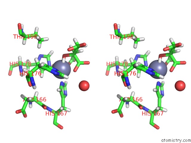
Stereo pair view
Mono view
Stereo pair view
A full contact list of Zinc with other atoms in the Zn binding
site number 1 of Crystal Structure of Zinc Binding Protein Zint in Complex with Citrate From E. Coli within 5.0Å range:
|
Zinc binding site 2 out of 2 in 5yxc
Go back to
Zinc binding site 2 out
of 2 in the Crystal Structure of Zinc Binding Protein Zint in Complex with Citrate From E. Coli

Mono view
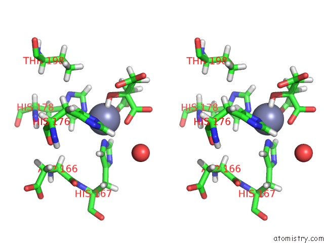
Stereo pair view
Mono view
Stereo pair view
A full contact list of Zinc with other atoms in the Zn binding
site number 2 of Crystal Structure of Zinc Binding Protein Zint in Complex with Citrate From E. Coli within 5.0Å range:
|
Reference:
J.Chen,
L.Wang,
F.Shang,
Y.Dong,
N.C.Ha,
K.H.Nam,
C.Quan,
Y.Xu.
Crystal Structure of E. Coli Zint with One Zinc-Binding Mode and Complexed with Citrate Biochem. Biophys. Res. V. 500 139 2018COMMUN..
ISSN: ESSN 1090-2104
PubMed: 29596824
DOI: 10.1016/J.BBRC.2018.03.192
Page generated: Mon Oct 28 16:28:42 2024
ISSN: ESSN 1090-2104
PubMed: 29596824
DOI: 10.1016/J.BBRC.2018.03.192
Last articles
Zn in 9MJ5Zn in 9HNW
Zn in 9G0L
Zn in 9FNE
Zn in 9DZN
Zn in 9E0I
Zn in 9D32
Zn in 9DAK
Zn in 8ZXC
Zn in 8ZUF