Zinc »
PDB 5xob-5y1s »
5xqz »
Zinc in PDB 5xqz: Structure of the MOB1-NDR2 Complex
Enzymatic activity of Structure of the MOB1-NDR2 Complex
All present enzymatic activity of Structure of the MOB1-NDR2 Complex:
2.7.11.1;
2.7.11.1;
Protein crystallography data
The structure of Structure of the MOB1-NDR2 Complex, PDB code: 5xqz
was solved by
G.Wu,
K.Lin,
with X-Ray Crystallography technique. A brief refinement statistics is given in the table below:
| Resolution Low / High (Å) | 50.00 / 2.10 |
| Space group | P 21 21 21 |
| Cell size a, b, c (Å), α, β, γ (°) | 57.519, 94.424, 102.200, 90.00, 90.00, 90.00 |
| R / Rfree (%) | 15.6 / 24.4 |
Zinc Binding Sites:
The binding sites of Zinc atom in the Structure of the MOB1-NDR2 Complex
(pdb code 5xqz). This binding sites where shown within
5.0 Angstroms radius around Zinc atom.
In total 2 binding sites of Zinc where determined in the Structure of the MOB1-NDR2 Complex, PDB code: 5xqz:
Jump to Zinc binding site number: 1; 2;
In total 2 binding sites of Zinc where determined in the Structure of the MOB1-NDR2 Complex, PDB code: 5xqz:
Jump to Zinc binding site number: 1; 2;
Zinc binding site 1 out of 2 in 5xqz
Go back to
Zinc binding site 1 out
of 2 in the Structure of the MOB1-NDR2 Complex

Mono view
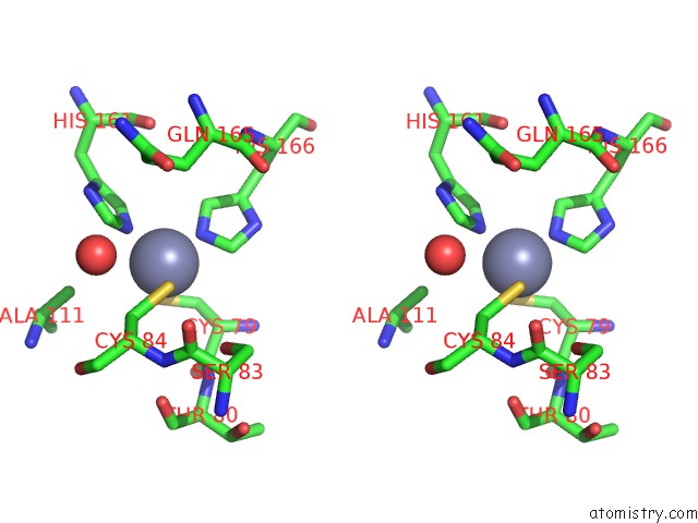
Stereo pair view
Mono view
Stereo pair view
A full contact list of Zinc with other atoms in the Zn binding
site number 1 of Structure of the MOB1-NDR2 Complex within 5.0Å range:
|
Zinc binding site 2 out of 2 in 5xqz
Go back to
Zinc binding site 2 out
of 2 in the Structure of the MOB1-NDR2 Complex
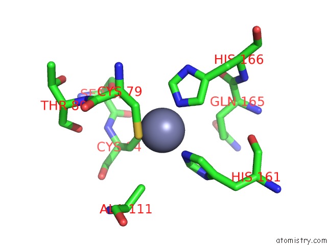
Mono view

Stereo pair view
Mono view
Stereo pair view
A full contact list of Zinc with other atoms in the Zn binding
site number 2 of Structure of the MOB1-NDR2 Complex within 5.0Å range:
|
Reference:
Y.Kulaberoglu,
K.Lin,
M.Holder,
Z.Gai,
M.Gomez,
B.Assefa Shifa,
M.Mavis,
L.Hoa,
A.A.D.Sharif,
C.Lujan,
E.S.J.Smith,
I.Bjedov,
N.Tapon,
G.Wu,
A.Hergovich.
Stable MOB1 Interaction with Hippo/Mst Is Not Essential For Development and Tissue Growth Control. Nat Commun V. 8 695 2017.
ISSN: ESSN 2041-1723
PubMed: 28947795
DOI: 10.1038/S41467-017-00795-Y
Page generated: Mon Oct 28 15:11:23 2024
ISSN: ESSN 2041-1723
PubMed: 28947795
DOI: 10.1038/S41467-017-00795-Y
Last articles
Zn in 9MJ5Zn in 9HNW
Zn in 9G0L
Zn in 9FNE
Zn in 9DZN
Zn in 9E0I
Zn in 9D32
Zn in 9DAK
Zn in 8ZXC
Zn in 8ZUF