Zinc »
PDB 5x6i-5xne »
5xfo »
Zinc in PDB 5xfo: Structure of the N-Terminal Domains of PHF1
Protein crystallography data
The structure of Structure of the N-Terminal Domains of PHF1, PDB code: 5xfo
was solved by
Z.Wang,
H.Li,
with X-Ray Crystallography technique. A brief refinement statistics is given in the table below:
| Resolution Low / High (Å) | 29.34 / 1.90 |
| Space group | P 21 21 21 |
| Cell size a, b, c (Å), α, β, γ (°) | 61.467, 66.772, 76.165, 90.00, 90.00, 90.00 |
| R / Rfree (%) | 17.7 / 22.7 |
Zinc Binding Sites:
The binding sites of Zinc atom in the Structure of the N-Terminal Domains of PHF1
(pdb code 5xfo). This binding sites where shown within
5.0 Angstroms radius around Zinc atom.
In total 4 binding sites of Zinc where determined in the Structure of the N-Terminal Domains of PHF1, PDB code: 5xfo:
Jump to Zinc binding site number: 1; 2; 3; 4;
In total 4 binding sites of Zinc where determined in the Structure of the N-Terminal Domains of PHF1, PDB code: 5xfo:
Jump to Zinc binding site number: 1; 2; 3; 4;
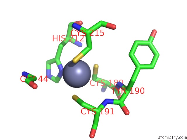
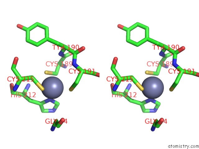
Zinc binding site 1 out of 4 in 5xfo
Go back to
Zinc binding site 1 out
of 4 in the Structure of the N-Terminal Domains of PHF1
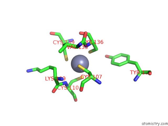
Mono view
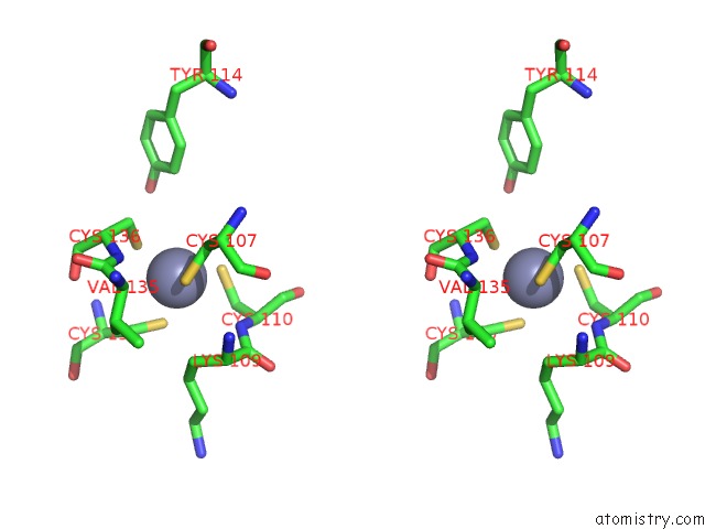
Stereo pair view
Mono view
Stereo pair view
A full contact list of Zinc with other atoms in the Zn binding
site number 1 of Structure of the N-Terminal Domains of PHF1 within 5.0Å range:
|
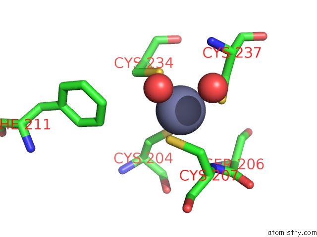
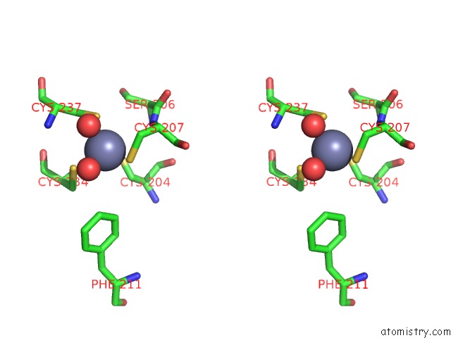
Zinc binding site 2 out of 4 in 5xfo
Go back to
Zinc binding site 2 out
of 4 in the Structure of the N-Terminal Domains of PHF1
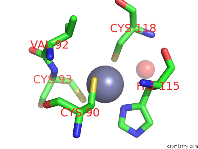
Mono view
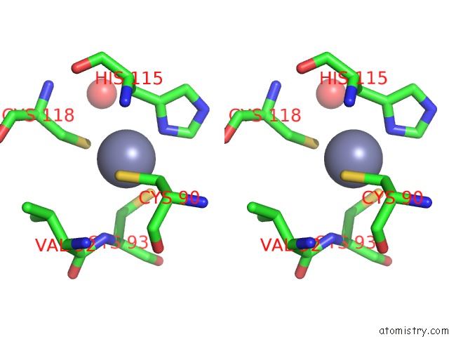
Stereo pair view
Mono view
Stereo pair view
A full contact list of Zinc with other atoms in the Zn binding
site number 2 of Structure of the N-Terminal Domains of PHF1 within 5.0Å range:
|
Zinc binding site 3 out of 4 in 5xfo
Go back to
Zinc binding site 3 out
of 4 in the Structure of the N-Terminal Domains of PHF1
Mono view
Stereo pair view
Mono view
Stereo pair view
A full contact list of Zinc with other atoms in the Zn binding
site number 3 of Structure of the N-Terminal Domains of PHF1 within 5.0Å range:
|
Zinc binding site 4 out of 4 in 5xfo
Go back to
Zinc binding site 4 out
of 4 in the Structure of the N-Terminal Domains of PHF1
Mono view
Stereo pair view
Mono view
Stereo pair view
A full contact list of Zinc with other atoms in the Zn binding
site number 4 of Structure of the N-Terminal Domains of PHF1 within 5.0Å range:
|
Reference:
H.Li,
R.Liefke,
J.Jiang,
J.V.Kurland,
W.Tian,
P.Deng,
W.Zhang,
Q.He,
D.J.Patel,
M.L.Bulyk,
Y.Shi,
Z.Wang.
Polycomb-Like Proteins Link the PRC2 Complex to Cpg Islands Nature V. 549 287 2017.
ISSN: ESSN 1476-4687
PubMed: 28869966
DOI: 10.1038/NATURE23881
Page generated: Mon Oct 28 14:58:18 2024
ISSN: ESSN 1476-4687
PubMed: 28869966
DOI: 10.1038/NATURE23881
Last articles
Zn in 9MJ5Zn in 9HNW
Zn in 9G0L
Zn in 9FNE
Zn in 9DZN
Zn in 9E0I
Zn in 9D32
Zn in 9DAK
Zn in 8ZXC
Zn in 8ZUF