Zinc »
PDB 5w0r-5w9q »
5w45 »
Zinc in PDB 5w45: Crystal Structure of APOBEC3H
Protein crystallography data
The structure of Crystal Structure of APOBEC3H, PDB code: 5w45
was solved by
F.Ito,
H.J.Yang,
X.Xiao,
S.X.Li,
A.Wolfe,
B.Zirkle,
V.Arutiunian,
X.J.Chen,
with X-Ray Crystallography technique. A brief refinement statistics is given in the table below:
| Resolution Low / High (Å) | 46.75 / 2.49 |
| Space group | P 1 21 1 |
| Cell size a, b, c (Å), α, β, γ (°) | 46.749, 65.006, 65.540, 90.00, 90.08, 90.00 |
| R / Rfree (%) | 21.1 / 23.5 |
Zinc Binding Sites:
The binding sites of Zinc atom in the Crystal Structure of APOBEC3H
(pdb code 5w45). This binding sites where shown within
5.0 Angstroms radius around Zinc atom.
In total 2 binding sites of Zinc where determined in the Crystal Structure of APOBEC3H, PDB code: 5w45:
Jump to Zinc binding site number: 1; 2;
In total 2 binding sites of Zinc where determined in the Crystal Structure of APOBEC3H, PDB code: 5w45:
Jump to Zinc binding site number: 1; 2;
Zinc binding site 1 out of 2 in 5w45
Go back to
Zinc binding site 1 out
of 2 in the Crystal Structure of APOBEC3H
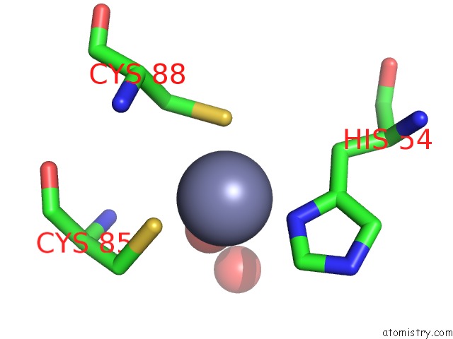
Mono view
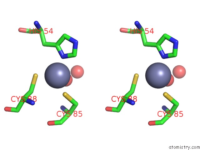
Stereo pair view
Mono view
Stereo pair view
A full contact list of Zinc with other atoms in the Zn binding
site number 1 of Crystal Structure of APOBEC3H within 5.0Å range:
|
Zinc binding site 2 out of 2 in 5w45
Go back to
Zinc binding site 2 out
of 2 in the Crystal Structure of APOBEC3H
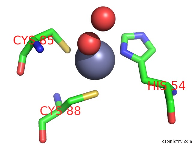
Mono view
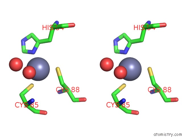
Stereo pair view
Mono view
Stereo pair view
A full contact list of Zinc with other atoms in the Zn binding
site number 2 of Crystal Structure of APOBEC3H within 5.0Å range:
|
Reference:
F.Ito,
H.Yang,
X.Xiao,
S.X.Li,
A.Wolfe,
B.Zirkle,
V.Arutiunian,
X.S.Chen.
Understanding the Structure, Multimerization, Subcellular Localization and Mc Selectivity of A Genomic Mutator and Anti-Hiv Factor APOBEC3H. Sci Rep V. 8 3763 2018.
ISSN: ESSN 2045-2322
PubMed: 29491387
DOI: 10.1038/S41598-018-21955-0
Page generated: Mon Oct 28 13:45:52 2024
ISSN: ESSN 2045-2322
PubMed: 29491387
DOI: 10.1038/S41598-018-21955-0
Last articles
Zn in 9MJ5Zn in 9HNW
Zn in 9G0L
Zn in 9FNE
Zn in 9DZN
Zn in 9E0I
Zn in 9D32
Zn in 9DAK
Zn in 8ZXC
Zn in 8ZUF