Zinc »
PDB 5w0r-5w9q »
5w2m »
Zinc in PDB 5w2m: APOBEC3F Catalytic Domain Complex with A Single-Stranded Dna
Protein crystallography data
The structure of APOBEC3F Catalytic Domain Complex with A Single-Stranded Dna, PDB code: 5w2m
was solved by
Y.Fang,
X.Xiao,
S.-X.Li,
A.Wolfe,
X.J.Chen,
with X-Ray Crystallography technique. A brief refinement statistics is given in the table below:
| Resolution Low / High (Å) | 50.00 / 3.70 |
| Space group | P 1 |
| Cell size a, b, c (Å), α, β, γ (°) | 68.194, 108.489, 108.549, 79.60, 71.80, 71.75 |
| R / Rfree (%) | 25 / 26.7 |
Zinc Binding Sites:
The binding sites of Zinc atom in the APOBEC3F Catalytic Domain Complex with A Single-Stranded Dna
(pdb code 5w2m). This binding sites where shown within
5.0 Angstroms radius around Zinc atom.
In total 4 binding sites of Zinc where determined in the APOBEC3F Catalytic Domain Complex with A Single-Stranded Dna, PDB code: 5w2m:
Jump to Zinc binding site number: 1; 2; 3; 4;
In total 4 binding sites of Zinc where determined in the APOBEC3F Catalytic Domain Complex with A Single-Stranded Dna, PDB code: 5w2m:
Jump to Zinc binding site number: 1; 2; 3; 4;
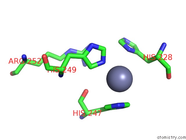
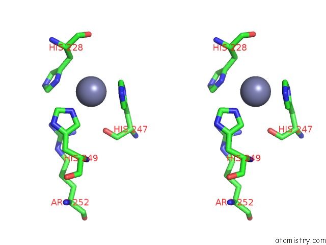
Zinc binding site 1 out of 4 in 5w2m
Go back to
Zinc binding site 1 out
of 4 in the APOBEC3F Catalytic Domain Complex with A Single-Stranded Dna
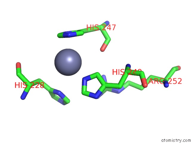
Mono view
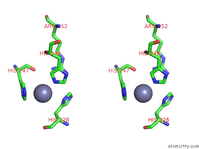
Stereo pair view
Mono view
Stereo pair view
A full contact list of Zinc with other atoms in the Zn binding
site number 1 of APOBEC3F Catalytic Domain Complex with A Single-Stranded Dna within 5.0Å range:
|
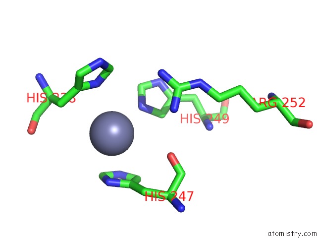
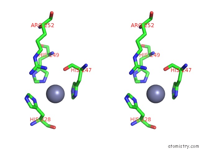
Zinc binding site 2 out of 4 in 5w2m
Go back to
Zinc binding site 2 out
of 4 in the APOBEC3F Catalytic Domain Complex with A Single-Stranded Dna
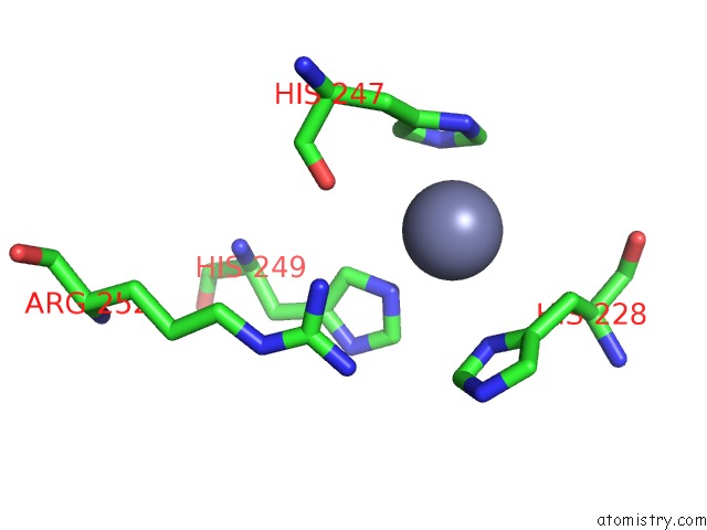
Mono view
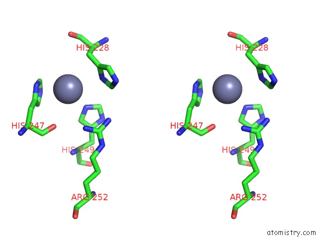
Stereo pair view
Mono view
Stereo pair view
A full contact list of Zinc with other atoms in the Zn binding
site number 2 of APOBEC3F Catalytic Domain Complex with A Single-Stranded Dna within 5.0Å range:
|
Zinc binding site 3 out of 4 in 5w2m
Go back to
Zinc binding site 3 out
of 4 in the APOBEC3F Catalytic Domain Complex with A Single-Stranded Dna
Mono view
Stereo pair view
Mono view
Stereo pair view
A full contact list of Zinc with other atoms in the Zn binding
site number 3 of APOBEC3F Catalytic Domain Complex with A Single-Stranded Dna within 5.0Å range:
|
Zinc binding site 4 out of 4 in 5w2m
Go back to
Zinc binding site 4 out
of 4 in the APOBEC3F Catalytic Domain Complex with A Single-Stranded Dna
Mono view
Stereo pair view
Mono view
Stereo pair view
A full contact list of Zinc with other atoms in the Zn binding
site number 4 of APOBEC3F Catalytic Domain Complex with A Single-Stranded Dna within 5.0Å range:
|
Reference:
Y.Fang,
X.Xiao,
S.X.Li,
A.Wolfe,
X.S.Chen.
Molecular Interactions of A Dna Modifying Enzyme APOBEC3F Catalytic Domain with A Single-Stranded Dna. J. Mol. Biol. V. 430 87 2018.
ISSN: ESSN 1089-8638
PubMed: 29191651
DOI: 10.1016/J.JMB.2017.11.007
Page generated: Mon Oct 28 13:41:35 2024
ISSN: ESSN 1089-8638
PubMed: 29191651
DOI: 10.1016/J.JMB.2017.11.007
Last articles
Zn in 9MJ5Zn in 9HNW
Zn in 9G0L
Zn in 9FNE
Zn in 9DZN
Zn in 9E0I
Zn in 9D32
Zn in 9DAK
Zn in 8ZXC
Zn in 8ZUF