Zinc »
PDB 5vmz-5vuq »
5vsw »
Zinc in PDB 5vsw: X-Ray Crystal Structure of Escherichia Coli Rna Polymerase and Dksa/Ppgpp Complex
Enzymatic activity of X-Ray Crystal Structure of Escherichia Coli Rna Polymerase and Dksa/Ppgpp Complex
All present enzymatic activity of X-Ray Crystal Structure of Escherichia Coli Rna Polymerase and Dksa/Ppgpp Complex:
2.7.7.6;
2.7.7.6;
Protein crystallography data
The structure of X-Ray Crystal Structure of Escherichia Coli Rna Polymerase and Dksa/Ppgpp Complex, PDB code: 5vsw
was solved by
K.S.Murakami,
V.Molodtsov,
with X-Ray Crystallography technique. A brief refinement statistics is given in the table below:
| Resolution Low / High (Å) | 46.82 / 4.30 |
| Space group | P 21 21 21 |
| Cell size a, b, c (Å), α, β, γ (°) | 187.274, 205.259, 311.313, 90.00, 90.00, 90.00 |
| R / Rfree (%) | 22.1 / 25.9 |
Other elements in 5vsw:
The structure of X-Ray Crystal Structure of Escherichia Coli Rna Polymerase and Dksa/Ppgpp Complex also contains other interesting chemical elements:
| Magnesium | (Mg) | 2 atoms |
Zinc Binding Sites:
The binding sites of Zinc atom in the X-Ray Crystal Structure of Escherichia Coli Rna Polymerase and Dksa/Ppgpp Complex
(pdb code 5vsw). This binding sites where shown within
5.0 Angstroms radius around Zinc atom.
In total 5 binding sites of Zinc where determined in the X-Ray Crystal Structure of Escherichia Coli Rna Polymerase and Dksa/Ppgpp Complex, PDB code: 5vsw:
Jump to Zinc binding site number: 1; 2; 3; 4; 5;
In total 5 binding sites of Zinc where determined in the X-Ray Crystal Structure of Escherichia Coli Rna Polymerase and Dksa/Ppgpp Complex, PDB code: 5vsw:
Jump to Zinc binding site number: 1; 2; 3; 4; 5;
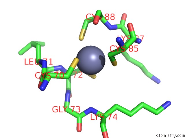
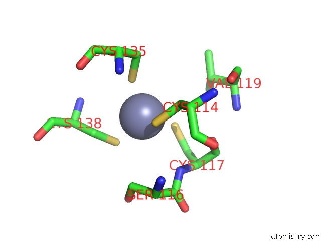
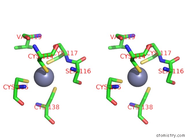
Zinc binding site 1 out of 5 in 5vsw
Go back to
Zinc binding site 1 out
of 5 in the X-Ray Crystal Structure of Escherichia Coli Rna Polymerase and Dksa/Ppgpp Complex
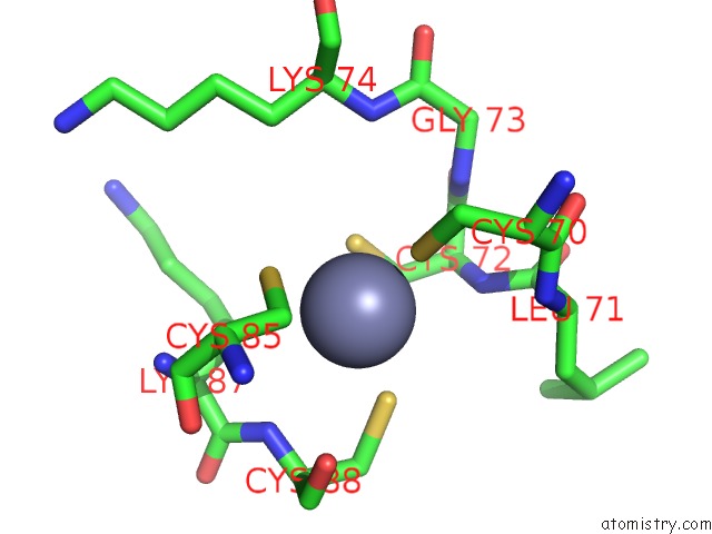
Mono view
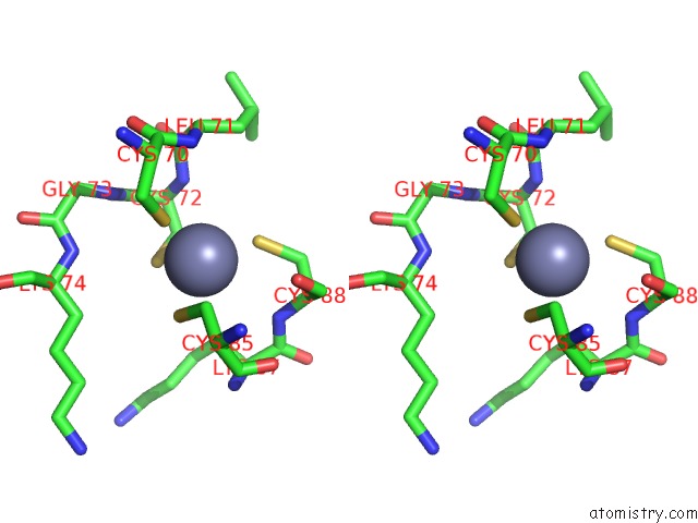
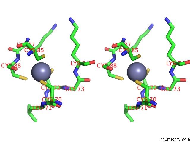
Stereo pair view
Mono view
Stereo pair view
A full contact list of Zinc with other atoms in the Zn binding
site number 1 of X-Ray Crystal Structure of Escherichia Coli Rna Polymerase and Dksa/Ppgpp Complex within 5.0Å range:
|
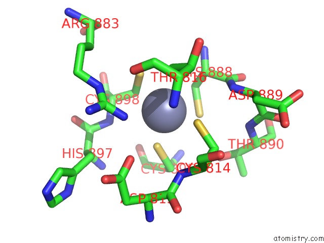
Zinc binding site 2 out of 5 in 5vsw
Go back to
Zinc binding site 2 out
of 5 in the X-Ray Crystal Structure of Escherichia Coli Rna Polymerase and Dksa/Ppgpp Complex
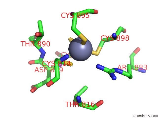
Mono view
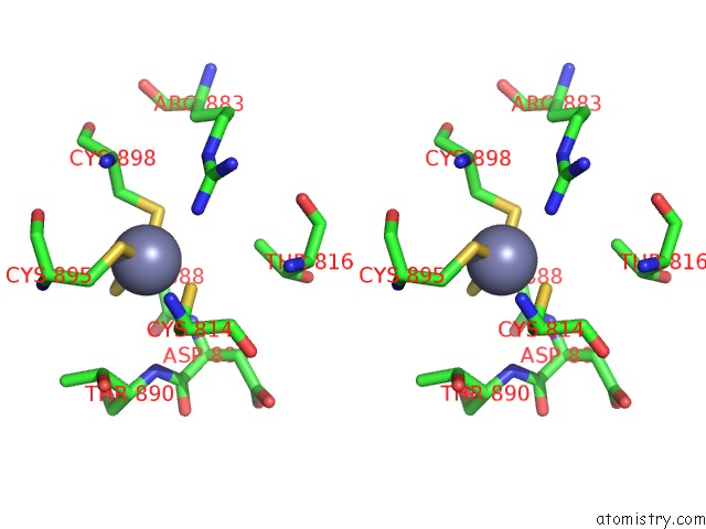
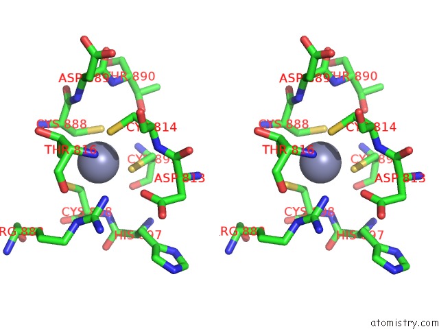
Stereo pair view
Mono view
Stereo pair view
A full contact list of Zinc with other atoms in the Zn binding
site number 2 of X-Ray Crystal Structure of Escherichia Coli Rna Polymerase and Dksa/Ppgpp Complex within 5.0Å range:
|
Zinc binding site 3 out of 5 in 5vsw
Go back to
Zinc binding site 3 out
of 5 in the X-Ray Crystal Structure of Escherichia Coli Rna Polymerase and Dksa/Ppgpp Complex
Mono view
Stereo pair view
Mono view
Stereo pair view
A full contact list of Zinc with other atoms in the Zn binding
site number 3 of X-Ray Crystal Structure of Escherichia Coli Rna Polymerase and Dksa/Ppgpp Complex within 5.0Å range:
|
Zinc binding site 4 out of 5 in 5vsw
Go back to
Zinc binding site 4 out
of 5 in the X-Ray Crystal Structure of Escherichia Coli Rna Polymerase and Dksa/Ppgpp Complex
Mono view
Stereo pair view
Mono view
Stereo pair view
A full contact list of Zinc with other atoms in the Zn binding
site number 4 of X-Ray Crystal Structure of Escherichia Coli Rna Polymerase and Dksa/Ppgpp Complex within 5.0Å range:
|
Zinc binding site 5 out of 5 in 5vsw
Go back to
Zinc binding site 5 out
of 5 in the X-Ray Crystal Structure of Escherichia Coli Rna Polymerase and Dksa/Ppgpp Complex
Mono view
Stereo pair view
Mono view
Stereo pair view
A full contact list of Zinc with other atoms in the Zn binding
site number 5 of X-Ray Crystal Structure of Escherichia Coli Rna Polymerase and Dksa/Ppgpp Complex within 5.0Å range:
|
Reference:
V.Molodtsov,
E.Sineva,
L.Zhang,
X.Huang,
M.Cashel,
S.E.Ades,
K.S.Murakami.
Allosteric Effector Ppgpp Potentiates the Inhibition of Transcript Initiation By Dksa. Mol. Cell V. 69 828 2018.
ISSN: ISSN 1097-4164
PubMed: 29478808
DOI: 10.1016/J.MOLCEL.2018.01.035
Page generated: Mon Oct 28 13:23:15 2024
ISSN: ISSN 1097-4164
PubMed: 29478808
DOI: 10.1016/J.MOLCEL.2018.01.035
Last articles
Zn in 9MJ5Zn in 9HNW
Zn in 9G0L
Zn in 9FNE
Zn in 9DZN
Zn in 9E0I
Zn in 9D32
Zn in 9DAK
Zn in 8ZXC
Zn in 8ZUF