Zinc »
PDB 5um3-5uqp »
5umf »
Zinc in PDB 5umf: Crystal Structure of A Ribulose-Phosphate 3-Epimerase From Neisseria Gonorrhoeae with Bound Phosphate
Enzymatic activity of Crystal Structure of A Ribulose-Phosphate 3-Epimerase From Neisseria Gonorrhoeae with Bound Phosphate
All present enzymatic activity of Crystal Structure of A Ribulose-Phosphate 3-Epimerase From Neisseria Gonorrhoeae with Bound Phosphate:
5.1.3.1;
5.1.3.1;
Protein crystallography data
The structure of Crystal Structure of A Ribulose-Phosphate 3-Epimerase From Neisseria Gonorrhoeae with Bound Phosphate, PDB code: 5umf
was solved by
Seattle Structural Genomics Center For Infectious Disease,
Seattlestructural Genomics Center For Infectious Disease (Ssgcid),
with X-Ray Crystallography technique. A brief refinement statistics is given in the table below:
| Resolution Low / High (Å) | 40.68 / 1.40 |
| Space group | C 1 2 1 |
| Cell size a, b, c (Å), α, β, γ (°) | 122.730, 70.900, 83.500, 90.00, 109.31, 90.00 |
| R / Rfree (%) | 14.8 / 16.5 |
Zinc Binding Sites:
The binding sites of Zinc atom in the Crystal Structure of A Ribulose-Phosphate 3-Epimerase From Neisseria Gonorrhoeae with Bound Phosphate
(pdb code 5umf). This binding sites where shown within
5.0 Angstroms radius around Zinc atom.
In total 3 binding sites of Zinc where determined in the Crystal Structure of A Ribulose-Phosphate 3-Epimerase From Neisseria Gonorrhoeae with Bound Phosphate, PDB code: 5umf:
Jump to Zinc binding site number: 1; 2; 3;
In total 3 binding sites of Zinc where determined in the Crystal Structure of A Ribulose-Phosphate 3-Epimerase From Neisseria Gonorrhoeae with Bound Phosphate, PDB code: 5umf:
Jump to Zinc binding site number: 1; 2; 3;
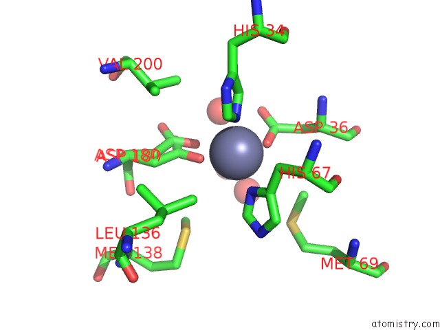
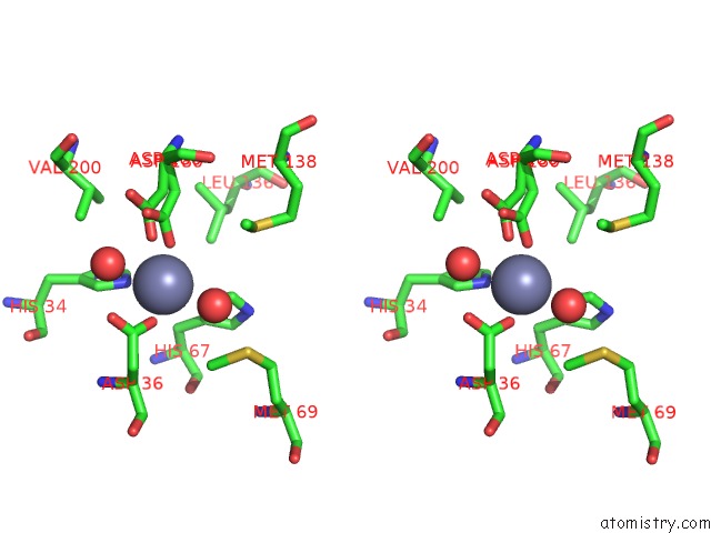
Zinc binding site 1 out of 3 in 5umf
Go back to
Zinc binding site 1 out
of 3 in the Crystal Structure of A Ribulose-Phosphate 3-Epimerase From Neisseria Gonorrhoeae with Bound Phosphate

Mono view
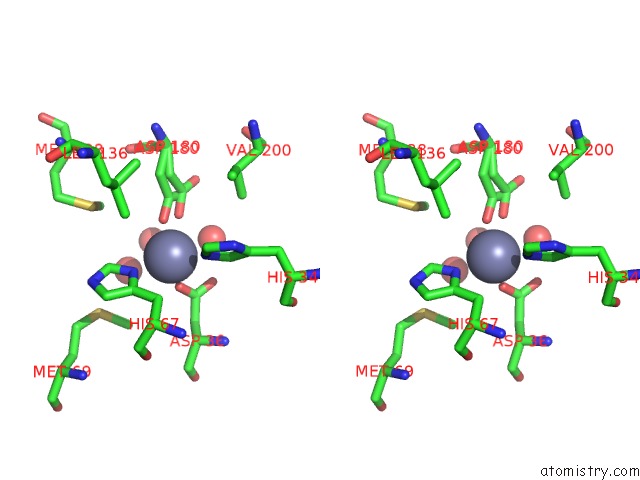
Stereo pair view
Mono view
Stereo pair view
A full contact list of Zinc with other atoms in the Zn binding
site number 1 of Crystal Structure of A Ribulose-Phosphate 3-Epimerase From Neisseria Gonorrhoeae with Bound Phosphate within 5.0Å range:
|
Zinc binding site 2 out of 3 in 5umf
Go back to
Zinc binding site 2 out
of 3 in the Crystal Structure of A Ribulose-Phosphate 3-Epimerase From Neisseria Gonorrhoeae with Bound Phosphate
Mono view
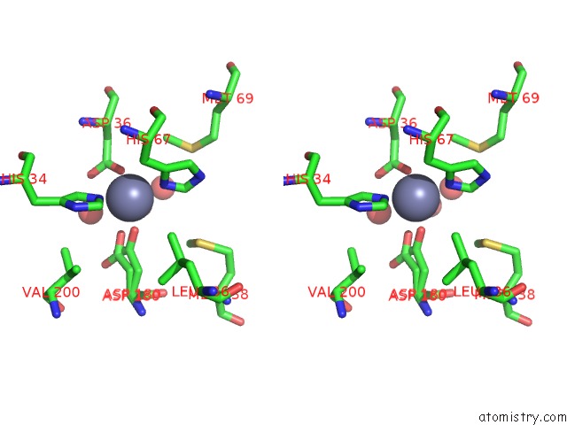
Stereo pair view
Mono view
Stereo pair view
A full contact list of Zinc with other atoms in the Zn binding
site number 2 of Crystal Structure of A Ribulose-Phosphate 3-Epimerase From Neisseria Gonorrhoeae with Bound Phosphate within 5.0Å range:
|
Zinc binding site 3 out of 3 in 5umf
Go back to
Zinc binding site 3 out
of 3 in the Crystal Structure of A Ribulose-Phosphate 3-Epimerase From Neisseria Gonorrhoeae with Bound Phosphate
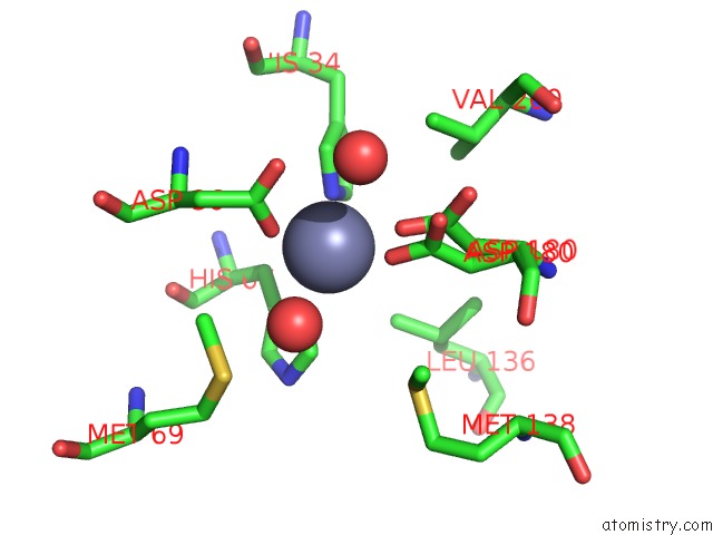
Mono view
Stereo pair view
Mono view
Stereo pair view
A full contact list of Zinc with other atoms in the Zn binding
site number 3 of Crystal Structure of A Ribulose-Phosphate 3-Epimerase From Neisseria Gonorrhoeae with Bound Phosphate within 5.0Å range:
|
Reference:
D.M.Dranow,
D.G.Conrady,
D.D.Lorimer,
T.E.Edwards.
Crystal Structure of A Ribulose-Phosphate 3-Epimerase From Neisseria Gonorrhoeae with Bound Phosphate To Be Published.
Page generated: Mon Oct 28 11:17:24 2024
Last articles
Al in 7NICAl in 7NIQ
Al in 7L07
Al in 7N77
Al in 7N73
Al in 7N72
Al in 7LVR
Al in 7KYB
Al in 7JL3
Al in 7KRO