Zinc »
PDB 5ucp-5uln »
5ufl »
Zinc in PDB 5ufl: Crystal Structure of A CIP2A Core Domain
Protein crystallography data
The structure of Crystal Structure of A CIP2A Core Domain, PDB code: 5ufl
was solved by
Z.Wang,
J.Wang,
Z.Rao,
W.Xu,
with X-Ray Crystallography technique. A brief refinement statistics is given in the table below:
| Resolution Low / High (Å) | 132.74 / 3.00 |
| Space group | P 65 |
| Cell size a, b, c (Å), α, β, γ (°) | 153.276, 153.276, 105.440, 90.00, 90.00, 120.00 |
| R / Rfree (%) | 25.6 / 28.1 |
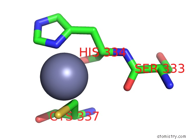
Zinc Binding Sites:
The binding sites of Zinc atom in the Crystal Structure of A CIP2A Core Domain
(pdb code 5ufl). This binding sites where shown within
5.0 Angstroms radius around Zinc atom.
In total only one binding site of Zinc was determined in the Crystal Structure of A CIP2A Core Domain, PDB code: 5ufl:
In total only one binding site of Zinc was determined in the Crystal Structure of A CIP2A Core Domain, PDB code: 5ufl:
Zinc binding site 1 out of 1 in 5ufl
Go back to
Zinc binding site 1 out
of 1 in the Crystal Structure of A CIP2A Core Domain
Mono view

Stereo pair view
Mono view
Stereo pair view
A full contact list of Zinc with other atoms in the Zn binding
site number 1 of Crystal Structure of A CIP2A Core Domain within 5.0Å range:
|
Reference:
J.Wang,
J.Okkeri,
K.Pavic,
Z.Wang,
O.Kauko,
T.Halonen,
G.Sarek,
P.M.Ojala,
Z.Rao,
W.Xu,
J.Westermarck.
Oncoprotein CIP2A Is Stabilized Via Interaction with Tumor Suppressor PP2A/B56. Embo Rep. V. 18 437 2017.
ISSN: ESSN 1469-3178
PubMed: 28174209
DOI: 10.15252/EMBR.201642788
Page generated: Mon Oct 28 09:30:42 2024
ISSN: ESSN 1469-3178
PubMed: 28174209
DOI: 10.15252/EMBR.201642788
Last articles
Zn in 9MJ5Zn in 9HNW
Zn in 9G0L
Zn in 9FNE
Zn in 9DZN
Zn in 9E0I
Zn in 9D32
Zn in 9DAK
Zn in 8ZXC
Zn in 8ZUF