Zinc »
PDB 5thi-5tt8 »
5thj »
Zinc in PDB 5thj: Crystal Structure of 2-Hydroxycyclohepta-2,4,6-Trien-1-One Bound to Human Carbonic Anhydrase 2
Enzymatic activity of Crystal Structure of 2-Hydroxycyclohepta-2,4,6-Trien-1-One Bound to Human Carbonic Anhydrase 2
All present enzymatic activity of Crystal Structure of 2-Hydroxycyclohepta-2,4,6-Trien-1-One Bound to Human Carbonic Anhydrase 2:
4.2.1.1;
4.2.1.1;
Protein crystallography data
The structure of Crystal Structure of 2-Hydroxycyclohepta-2,4,6-Trien-1-One Bound to Human Carbonic Anhydrase 2, PDB code: 5thj
was solved by
B.Dick,
S.Cohen,
with X-Ray Crystallography technique. A brief refinement statistics is given in the table below:
| Resolution Low / High (Å) | 40.00 / 1.50 |
| Space group | P 1 21 1 |
| Cell size a, b, c (Å), α, β, γ (°) | 42.283, 41.340, 71.848, 90.00, 104.00, 90.00 |
| R / Rfree (%) | 15.4 / 18.9 |
Other elements in 5thj:
The structure of Crystal Structure of 2-Hydroxycyclohepta-2,4,6-Trien-1-One Bound to Human Carbonic Anhydrase 2 also contains other interesting chemical elements:
| Mercury | (Hg) | 1 atom |
Zinc Binding Sites:
The binding sites of Zinc atom in the Crystal Structure of 2-Hydroxycyclohepta-2,4,6-Trien-1-One Bound to Human Carbonic Anhydrase 2
(pdb code 5thj). This binding sites where shown within
5.0 Angstroms radius around Zinc atom.
In total only one binding site of Zinc was determined in the Crystal Structure of 2-Hydroxycyclohepta-2,4,6-Trien-1-One Bound to Human Carbonic Anhydrase 2, PDB code: 5thj:
In total only one binding site of Zinc was determined in the Crystal Structure of 2-Hydroxycyclohepta-2,4,6-Trien-1-One Bound to Human Carbonic Anhydrase 2, PDB code: 5thj:
Zinc binding site 1 out of 1 in 5thj
Go back to
Zinc binding site 1 out
of 1 in the Crystal Structure of 2-Hydroxycyclohepta-2,4,6-Trien-1-One Bound to Human Carbonic Anhydrase 2
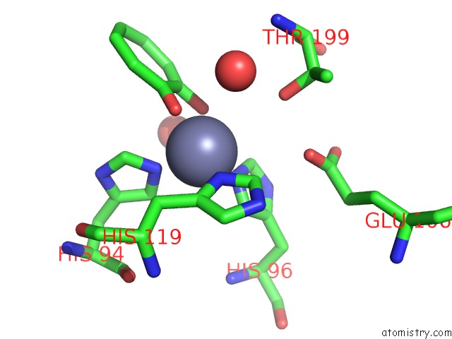
Mono view
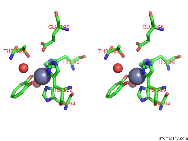
Stereo pair view
Mono view
Stereo pair view
A full contact list of Zinc with other atoms in the Zn binding
site number 1 of Crystal Structure of 2-Hydroxycyclohepta-2,4,6-Trien-1-One Bound to Human Carbonic Anhydrase 2 within 5.0Å range:
|
Reference:
B.L.Dick,
A.Patel,
J.A.Mccammon,
S.M.Cohen.
Effect of Donor Atom Identity on Metal-Binding Pharmacophore Coordination. J. Biol. Inorg. Chem. V. 22 605 2017.
ISSN: ESSN 1432-1327
PubMed: 28389830
DOI: 10.1007/S00775-017-1454-3
Page generated: Mon Oct 28 08:32:31 2024
ISSN: ESSN 1432-1327
PubMed: 28389830
DOI: 10.1007/S00775-017-1454-3
Last articles
Zn in 9MJ5Zn in 9HNW
Zn in 9G0L
Zn in 9FNE
Zn in 9DZN
Zn in 9E0I
Zn in 9D32
Zn in 9DAK
Zn in 8ZXC
Zn in 8ZUF