Zinc »
PDB 5t74-5th9 »
5tdc »
Zinc in PDB 5tdc: Crystal Structure of the Human Ubr-Box Domain From UBR1 in Complex with Monomethylated Arginine Peptide.
Protein crystallography data
The structure of Crystal Structure of the Human Ubr-Box Domain From UBR1 in Complex with Monomethylated Arginine Peptide., PDB code: 5tdc
was solved by
G.Kozlov,
J.Munoz-Escobar,
E.Matta-Camacho,
K.Gehring,
with X-Ray Crystallography technique. A brief refinement statistics is given in the table below:
| Resolution Low / High (Å) | 28.74 / 1.61 |
| Space group | P 21 21 21 |
| Cell size a, b, c (Å), α, β, γ (°) | 47.274, 49.036, 53.634, 90.00, 90.00, 90.00 |
| R / Rfree (%) | 14.9 / 18 |
Zinc Binding Sites:
The binding sites of Zinc atom in the Crystal Structure of the Human Ubr-Box Domain From UBR1 in Complex with Monomethylated Arginine Peptide.
(pdb code 5tdc). This binding sites where shown within
5.0 Angstroms radius around Zinc atom.
In total 6 binding sites of Zinc where determined in the Crystal Structure of the Human Ubr-Box Domain From UBR1 in Complex with Monomethylated Arginine Peptide., PDB code: 5tdc:
Jump to Zinc binding site number: 1; 2; 3; 4; 5; 6;
In total 6 binding sites of Zinc where determined in the Crystal Structure of the Human Ubr-Box Domain From UBR1 in Complex with Monomethylated Arginine Peptide., PDB code: 5tdc:
Jump to Zinc binding site number: 1; 2; 3; 4; 5; 6;
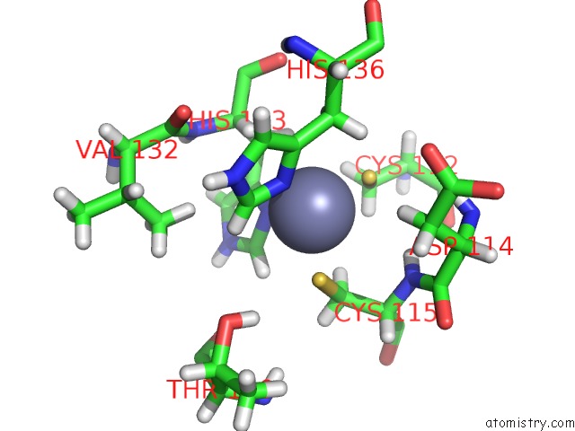
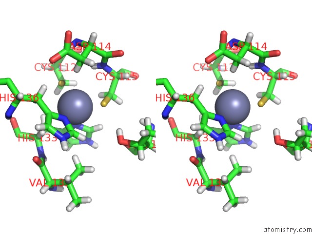
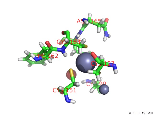
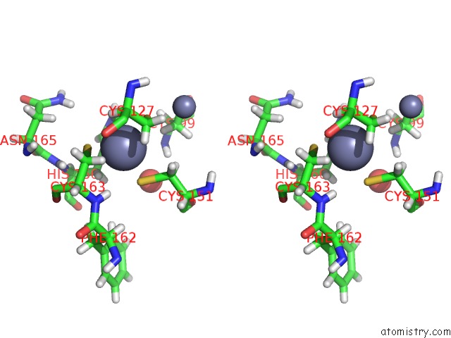
Zinc binding site 1 out of 6 in 5tdc
Go back to
Zinc binding site 1 out
of 6 in the Crystal Structure of the Human Ubr-Box Domain From UBR1 in Complex with Monomethylated Arginine Peptide.
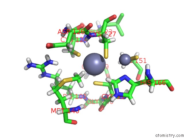
Mono view
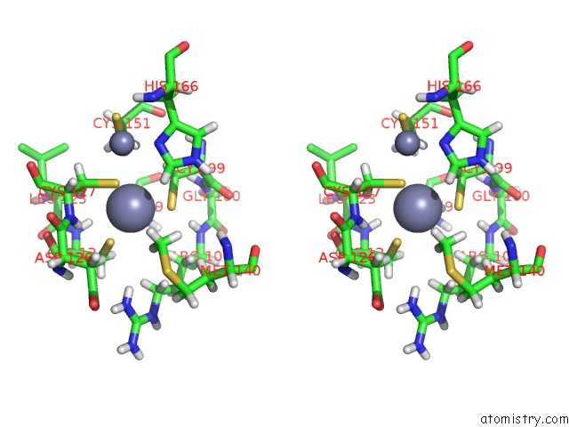
Stereo pair view
Mono view
Stereo pair view
A full contact list of Zinc with other atoms in the Zn binding
site number 1 of Crystal Structure of the Human Ubr-Box Domain From UBR1 in Complex with Monomethylated Arginine Peptide. within 5.0Å range:
|
Zinc binding site 2 out of 6 in 5tdc
Go back to
Zinc binding site 2 out
of 6 in the Crystal Structure of the Human Ubr-Box Domain From UBR1 in Complex with Monomethylated Arginine Peptide.
Mono view
Stereo pair view
Mono view
Stereo pair view
A full contact list of Zinc with other atoms in the Zn binding
site number 2 of Crystal Structure of the Human Ubr-Box Domain From UBR1 in Complex with Monomethylated Arginine Peptide. within 5.0Å range:
|
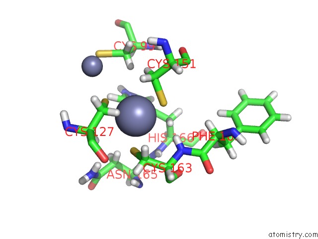
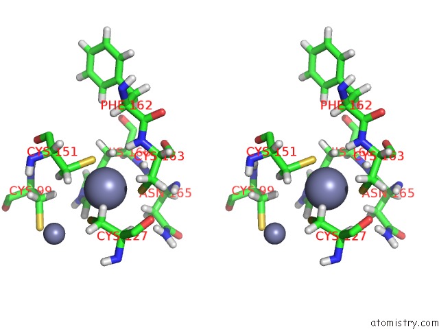
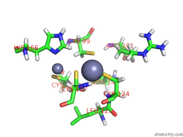
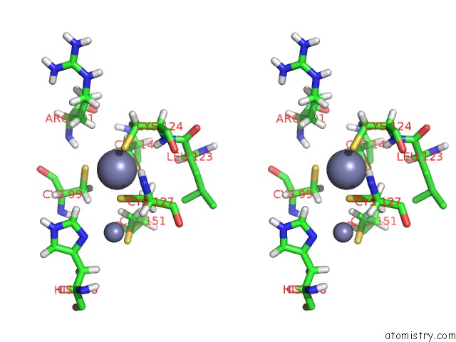
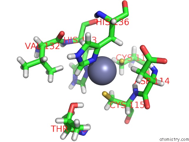
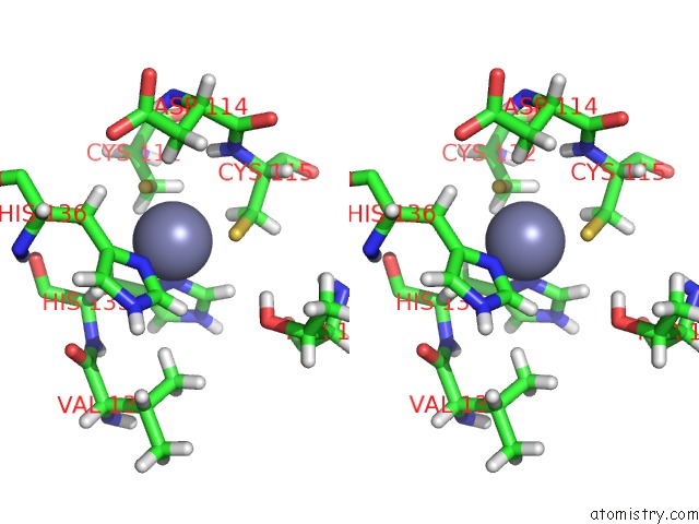
Zinc binding site 3 out of 6 in 5tdc
Go back to
Zinc binding site 3 out
of 6 in the Crystal Structure of the Human Ubr-Box Domain From UBR1 in Complex with Monomethylated Arginine Peptide.
Mono view
Stereo pair view
Mono view
Stereo pair view
A full contact list of Zinc with other atoms in the Zn binding
site number 3 of Crystal Structure of the Human Ubr-Box Domain From UBR1 in Complex with Monomethylated Arginine Peptide. within 5.0Å range:
|
Zinc binding site 4 out of 6 in 5tdc
Go back to
Zinc binding site 4 out
of 6 in the Crystal Structure of the Human Ubr-Box Domain From UBR1 in Complex with Monomethylated Arginine Peptide.
Mono view
Stereo pair view
Mono view
Stereo pair view
A full contact list of Zinc with other atoms in the Zn binding
site number 4 of Crystal Structure of the Human Ubr-Box Domain From UBR1 in Complex with Monomethylated Arginine Peptide. within 5.0Å range:
|
Zinc binding site 5 out of 6 in 5tdc
Go back to
Zinc binding site 5 out
of 6 in the Crystal Structure of the Human Ubr-Box Domain From UBR1 in Complex with Monomethylated Arginine Peptide.
Mono view
Stereo pair view
Mono view
Stereo pair view
A full contact list of Zinc with other atoms in the Zn binding
site number 5 of Crystal Structure of the Human Ubr-Box Domain From UBR1 in Complex with Monomethylated Arginine Peptide. within 5.0Å range:
|
Zinc binding site 6 out of 6 in 5tdc
Go back to
Zinc binding site 6 out
of 6 in the Crystal Structure of the Human Ubr-Box Domain From UBR1 in Complex with Monomethylated Arginine Peptide.
Mono view
Stereo pair view
Mono view
Stereo pair view
A full contact list of Zinc with other atoms in the Zn binding
site number 6 of Crystal Structure of the Human Ubr-Box Domain From UBR1 in Complex with Monomethylated Arginine Peptide. within 5.0Å range:
|
Reference:
J.Munoz-Escobar,
E.Matta-Camacho,
C.Cho,
G.Kozlov,
K.Gehring.
Bound Waters Mediate Binding of Diverse Substrates to A Ubiquitin Ligase. Structure V. 25 719 2017.
ISSN: ISSN 1878-4186
PubMed: 28392261
DOI: 10.1016/J.STR.2017.03.004
Page generated: Mon Oct 28 08:26:58 2024
ISSN: ISSN 1878-4186
PubMed: 28392261
DOI: 10.1016/J.STR.2017.03.004
Last articles
Zn in 9MJ5Zn in 9HNW
Zn in 9G0L
Zn in 9FNE
Zn in 9DZN
Zn in 9E0I
Zn in 9D32
Zn in 9DAK
Zn in 8ZXC
Zn in 8ZUF