Zinc »
PDB 5t74-5th9 »
5tab »
Zinc in PDB 5tab: Crystal Structure of the Phd Finger of PHF20
Protein crystallography data
The structure of Crystal Structure of the Phd Finger of PHF20, PDB code: 5tab
was solved by
B.J.Klein,
T.G.Kutateladze,
with X-Ray Crystallography technique. A brief refinement statistics is given in the table below:
| Resolution Low / High (Å) | 31.69 / 1.25 |
| Space group | P 61 |
| Cell size a, b, c (Å), α, β, γ (°) | 46.739, 46.739, 50.928, 90.00, 90.00, 120.00 |
| R / Rfree (%) | 11.8 / 14.4 |
Zinc Binding Sites:
The binding sites of Zinc atom in the Crystal Structure of the Phd Finger of PHF20
(pdb code 5tab). This binding sites where shown within
5.0 Angstroms radius around Zinc atom.
In total 2 binding sites of Zinc where determined in the Crystal Structure of the Phd Finger of PHF20, PDB code: 5tab:
Jump to Zinc binding site number: 1; 2;
In total 2 binding sites of Zinc where determined in the Crystal Structure of the Phd Finger of PHF20, PDB code: 5tab:
Jump to Zinc binding site number: 1; 2;
Zinc binding site 1 out of 2 in 5tab
Go back to
Zinc binding site 1 out
of 2 in the Crystal Structure of the Phd Finger of PHF20
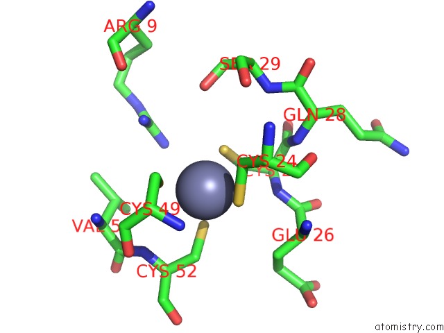
Mono view
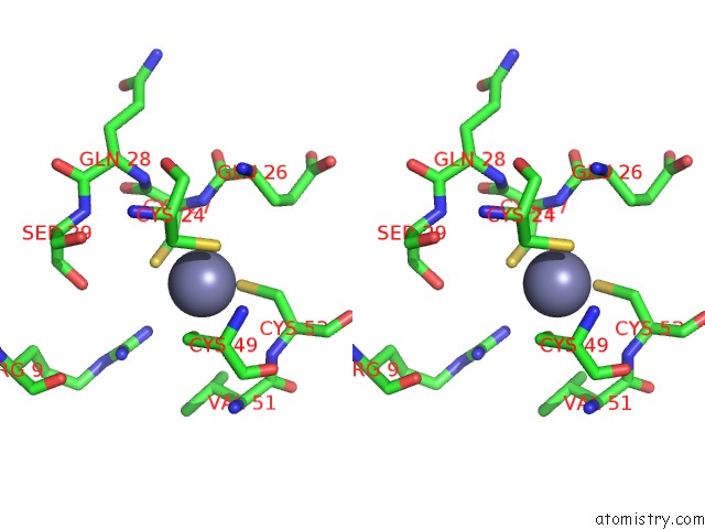
Stereo pair view
Mono view
Stereo pair view
A full contact list of Zinc with other atoms in the Zn binding
site number 1 of Crystal Structure of the Phd Finger of PHF20 within 5.0Å range:
|
Zinc binding site 2 out of 2 in 5tab
Go back to
Zinc binding site 2 out
of 2 in the Crystal Structure of the Phd Finger of PHF20
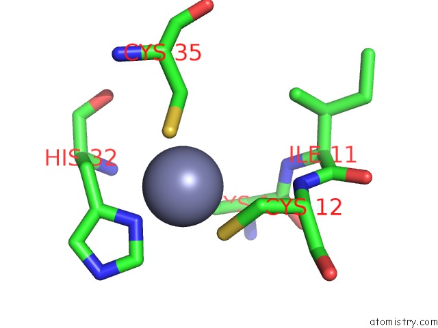
Mono view
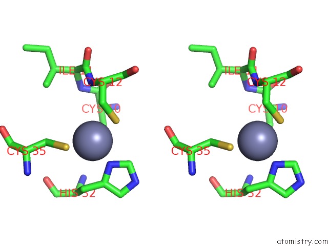
Stereo pair view
Mono view
Stereo pair view
A full contact list of Zinc with other atoms in the Zn binding
site number 2 of Crystal Structure of the Phd Finger of PHF20 within 5.0Å range:
|
Reference:
B.J.Klein,
X.Wang,
G.Cui,
C.Yuan,
M.V.Botuyan,
K.Lin,
Y.Lu,
X.Wang,
Y.Zhao,
C.J.Bruns,
G.Mer,
X.Shi,
T.G.Kutateladze.
PHF20 Readers Link Methylation of Histone H3K4 and P53 with H4K16 Acetylation. Cell Rep V. 17 1158 2016.
ISSN: ESSN 2211-1247
PubMed: 27760318
DOI: 10.1016/J.CELREP.2016.09.056
Page generated: Mon Oct 28 08:22:59 2024
ISSN: ESSN 2211-1247
PubMed: 27760318
DOI: 10.1016/J.CELREP.2016.09.056
Last articles
Zn in 9MJ5Zn in 9HNW
Zn in 9G0L
Zn in 9FNE
Zn in 9DZN
Zn in 9E0I
Zn in 9D32
Zn in 9DAK
Zn in 8ZXC
Zn in 8ZUF