Zinc »
PDB 5svc-5t72 »
5syt »
Zinc in PDB 5syt: Crystal Structure of ZMPSTE24
Enzymatic activity of Crystal Structure of ZMPSTE24
All present enzymatic activity of Crystal Structure of ZMPSTE24:
3.4.24.84;
3.4.24.84;
Protein crystallography data
The structure of Crystal Structure of ZMPSTE24, PDB code: 5syt
was solved by
K.Clark,
J.L.Jenkins,
N.Fedoriw,
M.E.Dumont,
Membrane Protein Structuralbiology Consortium (Mpsbc),
with X-Ray Crystallography technique. A brief refinement statistics is given in the table below:
| Resolution Low / High (Å) | 33.60 / 2.00 |
| Space group | C 1 2 1 |
| Cell size a, b, c (Å), α, β, γ (°) | 149.455, 84.560, 76.885, 90.00, 119.07, 90.00 |
| R / Rfree (%) | 21.8 / 24.9 |
Zinc Binding Sites:
The binding sites of Zinc atom in the Crystal Structure of ZMPSTE24
(pdb code 5syt). This binding sites where shown within
5.0 Angstroms radius around Zinc atom.
In total only one binding site of Zinc was determined in the Crystal Structure of ZMPSTE24, PDB code: 5syt:
In total only one binding site of Zinc was determined in the Crystal Structure of ZMPSTE24, PDB code: 5syt:
Zinc binding site 1 out of 1 in 5syt
Go back to
Zinc binding site 1 out
of 1 in the Crystal Structure of ZMPSTE24
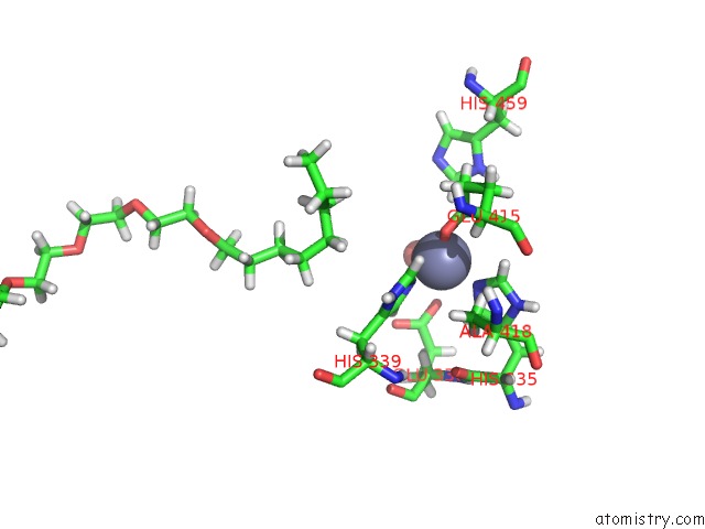
Mono view
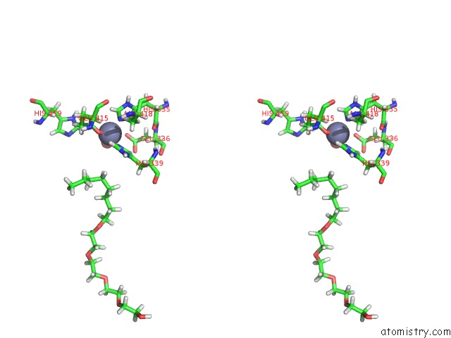
Stereo pair view
Mono view
Stereo pair view
A full contact list of Zinc with other atoms in the Zn binding
site number 1 of Crystal Structure of ZMPSTE24 within 5.0Å range:
|
Reference:
K.M.Clark,
J.L.Jenkins,
N.Fedoriw,
M.E.Dumont.
Human Caax Protease ZMPSTE24 Expressed in Yeast: Structure and Inhibition By Hiv Protease Inhibitors. Protein Sci. V. 26 242 2017.
ISSN: ESSN 1469-896X
PubMed: 27774687
DOI: 10.1002/PRO.3074
Page generated: Mon Oct 28 08:08:18 2024
ISSN: ESSN 1469-896X
PubMed: 27774687
DOI: 10.1002/PRO.3074
Last articles
Zn in 9MJ5Zn in 9HNW
Zn in 9G0L
Zn in 9FNE
Zn in 9DZN
Zn in 9E0I
Zn in 9D32
Zn in 9DAK
Zn in 8ZXC
Zn in 8ZUF