Zinc »
PDB 5o2f-5ofb »
5o2f »
Zinc in PDB 5o2f: Crystal Structure of Ndm-1 in Complex with Hydrolyzed Ampicillin - New Refinement
Enzymatic activity of Crystal Structure of Ndm-1 in Complex with Hydrolyzed Ampicillin - New Refinement
All present enzymatic activity of Crystal Structure of Ndm-1 in Complex with Hydrolyzed Ampicillin - New Refinement:
3.5.2.6;
3.5.2.6;
Protein crystallography data
The structure of Crystal Structure of Ndm-1 in Complex with Hydrolyzed Ampicillin - New Refinement, PDB code: 5o2f
was solved by
J.E.Raczynska,
I.G.Shabalin,
M.Jaskolski,
W.Minor,
A.Wlodawer,
with X-Ray Crystallography technique. A brief refinement statistics is given in the table below:
| Resolution Low / High (Å) | 38.79 / 2.01 |
| Space group | P 21 21 21 |
| Cell size a, b, c (Å), α, β, γ (°) | 38.620, 77.570, 132.310, 90.00, 90.00, 90.00 |
| R / Rfree (%) | 13.2 / 18.2 |
Other elements in 5o2f:
The structure of Crystal Structure of Ndm-1 in Complex with Hydrolyzed Ampicillin - New Refinement also contains other interesting chemical elements:
| Chlorine | (Cl) | 1 atom |
Zinc Binding Sites:
The binding sites of Zinc atom in the Crystal Structure of Ndm-1 in Complex with Hydrolyzed Ampicillin - New Refinement
(pdb code 5o2f). This binding sites where shown within
5.0 Angstroms radius around Zinc atom.
In total 4 binding sites of Zinc where determined in the Crystal Structure of Ndm-1 in Complex with Hydrolyzed Ampicillin - New Refinement, PDB code: 5o2f:
Jump to Zinc binding site number: 1; 2; 3; 4;
In total 4 binding sites of Zinc where determined in the Crystal Structure of Ndm-1 in Complex with Hydrolyzed Ampicillin - New Refinement, PDB code: 5o2f:
Jump to Zinc binding site number: 1; 2; 3; 4;
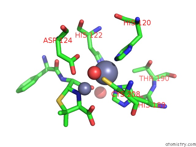
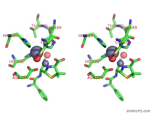
Zinc binding site 1 out of 4 in 5o2f
Go back to
Zinc binding site 1 out
of 4 in the Crystal Structure of Ndm-1 in Complex with Hydrolyzed Ampicillin - New Refinement
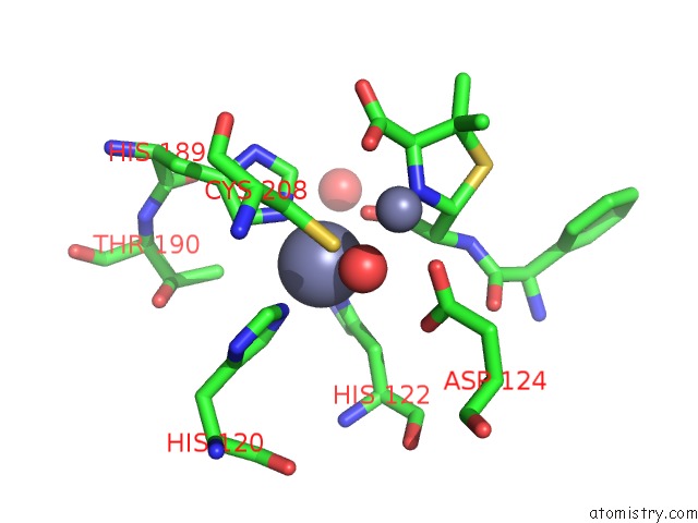
Mono view
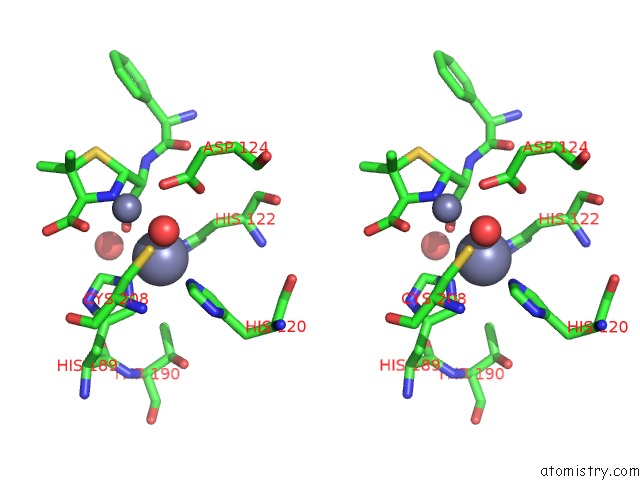
Stereo pair view
Mono view
Stereo pair view
A full contact list of Zinc with other atoms in the Zn binding
site number 1 of Crystal Structure of Ndm-1 in Complex with Hydrolyzed Ampicillin - New Refinement within 5.0Å range:
|
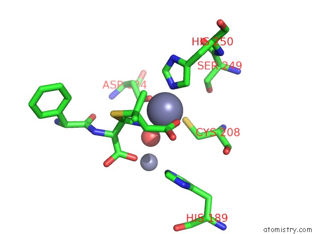
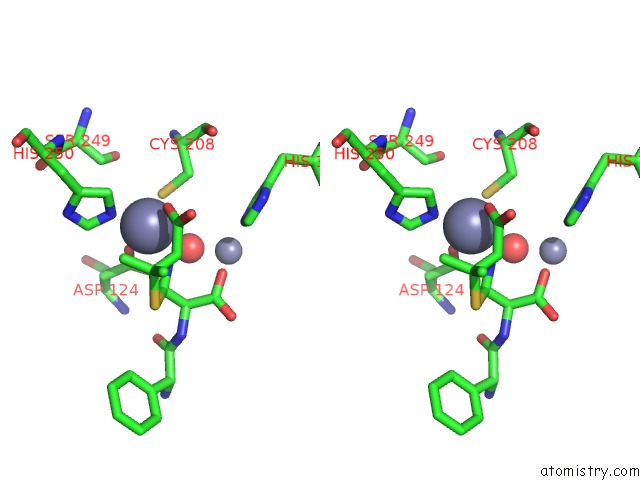
Zinc binding site 2 out of 4 in 5o2f
Go back to
Zinc binding site 2 out
of 4 in the Crystal Structure of Ndm-1 in Complex with Hydrolyzed Ampicillin - New Refinement
Mono view
Stereo pair view
Mono view
Stereo pair view
A full contact list of Zinc with other atoms in the Zn binding
site number 2 of Crystal Structure of Ndm-1 in Complex with Hydrolyzed Ampicillin - New Refinement within 5.0Å range:
|
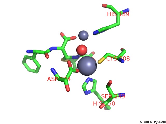
Zinc binding site 3 out of 4 in 5o2f
Go back to
Zinc binding site 3 out
of 4 in the Crystal Structure of Ndm-1 in Complex with Hydrolyzed Ampicillin - New Refinement
Mono view
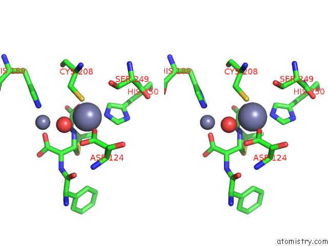
Stereo pair view
Mono view
Stereo pair view
A full contact list of Zinc with other atoms in the Zn binding
site number 3 of Crystal Structure of Ndm-1 in Complex with Hydrolyzed Ampicillin - New Refinement within 5.0Å range:
|
Zinc binding site 4 out of 4 in 5o2f
Go back to
Zinc binding site 4 out
of 4 in the Crystal Structure of Ndm-1 in Complex with Hydrolyzed Ampicillin - New Refinement
Mono view
Stereo pair view
Mono view
Stereo pair view
A full contact list of Zinc with other atoms in the Zn binding
site number 4 of Crystal Structure of Ndm-1 in Complex with Hydrolyzed Ampicillin - New Refinement within 5.0Å range:
|
Reference:
J.E.Raczynska,
I.G.Shabalin,
W.Minor,
A.Wlodawer,
M.Jaskolski.
A Close Look Onto Structural Models and Primary Ligands of Metallo-Beta-Lactamases. Drug Resist. Updat. V. 40 1 2018.
ISSN: ESSN 1532-2084
PubMed: 30466711
DOI: 10.1016/J.DRUP.2018.08.001
Page generated: Sun Oct 27 23:12:48 2024
ISSN: ESSN 1532-2084
PubMed: 30466711
DOI: 10.1016/J.DRUP.2018.08.001
Last articles
Zn in 9MJ5Zn in 9HNW
Zn in 9G0L
Zn in 9FNE
Zn in 9DZN
Zn in 9E0I
Zn in 9D32
Zn in 9DAK
Zn in 8ZXC
Zn in 8ZUF