Zinc »
PDB 5n38-5nel »
5nc9 »
Zinc in PDB 5nc9: Crystal Structure of the Polysaccharide Deacetylase BC1974 From Bacillus Cereus in Complex with (2S)-2,6-Diamino-N-Hydroxyhexanamide
Protein crystallography data
The structure of Crystal Structure of the Polysaccharide Deacetylase BC1974 From Bacillus Cereus in Complex with (2S)-2,6-Diamino-N-Hydroxyhexanamide, PDB code: 5nc9
was solved by
P.Giastas,
A.Andreou,
E.E.Eliopoulos,
with X-Ray Crystallography technique. A brief refinement statistics is given in the table below:
| Resolution Low / High (Å) | 48.61 / 2.44 |
| Space group | P 1 21 1 |
| Cell size a, b, c (Å), α, β, γ (°) | 49.767, 117.835, 99.059, 90.00, 102.38, 90.00 |
| R / Rfree (%) | 22 / 27.9 |
Zinc Binding Sites:
The binding sites of Zinc atom in the Crystal Structure of the Polysaccharide Deacetylase BC1974 From Bacillus Cereus in Complex with (2S)-2,6-Diamino-N-Hydroxyhexanamide
(pdb code 5nc9). This binding sites where shown within
5.0 Angstroms radius around Zinc atom.
In total 4 binding sites of Zinc where determined in the Crystal Structure of the Polysaccharide Deacetylase BC1974 From Bacillus Cereus in Complex with (2S)-2,6-Diamino-N-Hydroxyhexanamide, PDB code: 5nc9:
Jump to Zinc binding site number: 1; 2; 3; 4;
In total 4 binding sites of Zinc where determined in the Crystal Structure of the Polysaccharide Deacetylase BC1974 From Bacillus Cereus in Complex with (2S)-2,6-Diamino-N-Hydroxyhexanamide, PDB code: 5nc9:
Jump to Zinc binding site number: 1; 2; 3; 4;
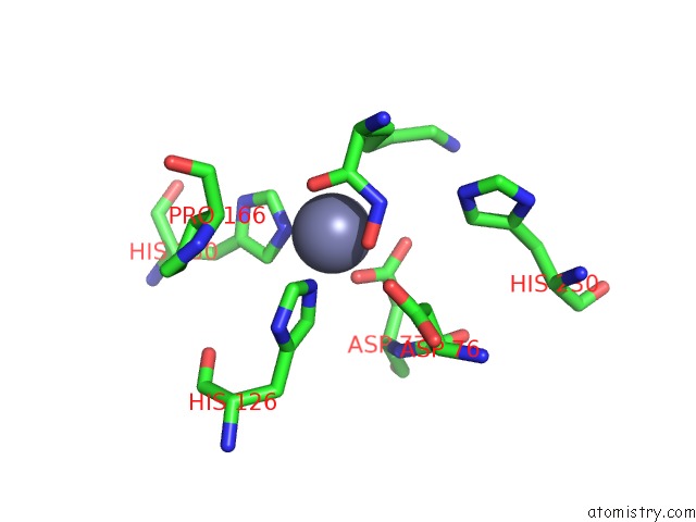
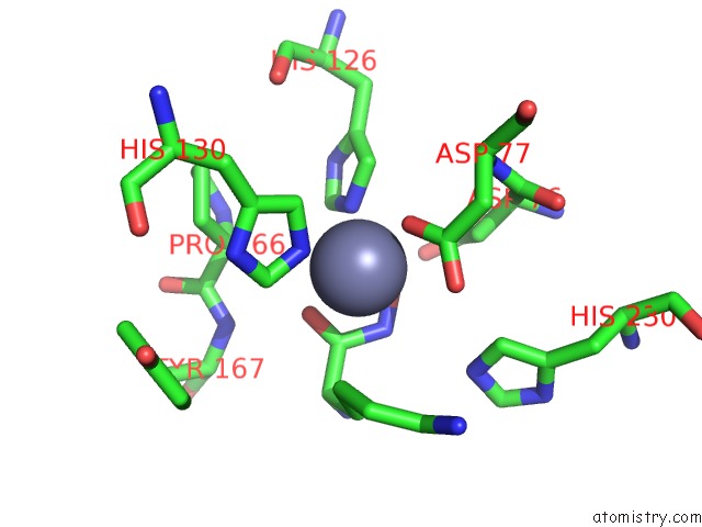
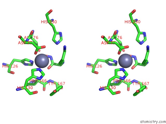
Zinc binding site 1 out of 4 in 5nc9
Go back to
Zinc binding site 1 out
of 4 in the Crystal Structure of the Polysaccharide Deacetylase BC1974 From Bacillus Cereus in Complex with (2S)-2,6-Diamino-N-Hydroxyhexanamide
Mono view
Stereo pair view
Mono view
Stereo pair view
A full contact list of Zinc with other atoms in the Zn binding
site number 1 of Crystal Structure of the Polysaccharide Deacetylase BC1974 From Bacillus Cereus in Complex with (2S)-2,6-Diamino-N-Hydroxyhexanamide within 5.0Å range:
|
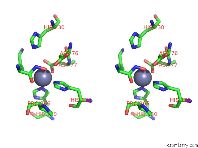
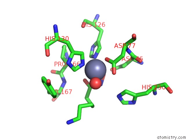
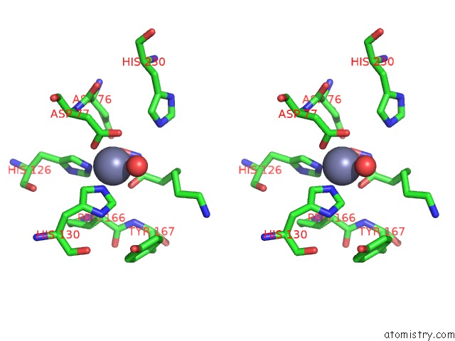
Zinc binding site 2 out of 4 in 5nc9
Go back to
Zinc binding site 2 out
of 4 in the Crystal Structure of the Polysaccharide Deacetylase BC1974 From Bacillus Cereus in Complex with (2S)-2,6-Diamino-N-Hydroxyhexanamide
Mono view
Stereo pair view
Mono view
Stereo pair view
A full contact list of Zinc with other atoms in the Zn binding
site number 2 of Crystal Structure of the Polysaccharide Deacetylase BC1974 From Bacillus Cereus in Complex with (2S)-2,6-Diamino-N-Hydroxyhexanamide within 5.0Å range:
|
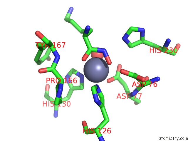
Zinc binding site 3 out of 4 in 5nc9
Go back to
Zinc binding site 3 out
of 4 in the Crystal Structure of the Polysaccharide Deacetylase BC1974 From Bacillus Cereus in Complex with (2S)-2,6-Diamino-N-Hydroxyhexanamide
Mono view
Stereo pair view
Mono view
Stereo pair view
A full contact list of Zinc with other atoms in the Zn binding
site number 3 of Crystal Structure of the Polysaccharide Deacetylase BC1974 From Bacillus Cereus in Complex with (2S)-2,6-Diamino-N-Hydroxyhexanamide within 5.0Å range:
|
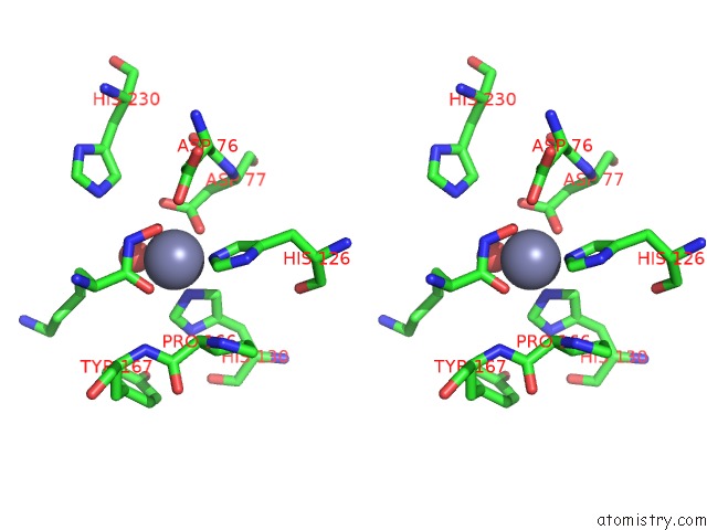
Zinc binding site 4 out of 4 in 5nc9
Go back to
Zinc binding site 4 out
of 4 in the Crystal Structure of the Polysaccharide Deacetylase BC1974 From Bacillus Cereus in Complex with (2S)-2,6-Diamino-N-Hydroxyhexanamide
Mono view
Stereo pair view
Mono view
Stereo pair view
A full contact list of Zinc with other atoms in the Zn binding
site number 4 of Crystal Structure of the Polysaccharide Deacetylase BC1974 From Bacillus Cereus in Complex with (2S)-2,6-Diamino-N-Hydroxyhexanamide within 5.0Å range:
|
Reference:
P.Giastas,
A.Andreou,
A.Papakyriakou,
D.Koutsioulis,
S.Balomenou,
S.J.Tzartos,
V.Bouriotis,
E.E.Eliopoulos.
Structures of the Peptidoglycan N-Acetylglucosamine Deacetylase BC1974 and Its Complexes with Zinc Metalloenzyme Inhibitors. Biochemistry V. 57 753 2018.
ISSN: ISSN 1520-4995
PubMed: 29257674
DOI: 10.1021/ACS.BIOCHEM.7B00919
Page generated: Thu Aug 21 05:32:39 2025
ISSN: ISSN 1520-4995
PubMed: 29257674
DOI: 10.1021/ACS.BIOCHEM.7B00919
Last articles
Zn in 6GNIZn in 6GM9
Zn in 6GJW
Zn in 6GL1
Zn in 6GKH
Zn in 6GKM
Zn in 6GHC
Zn in 6GJK
Zn in 6GJZ
Zn in 6GHS