Zinc »
PDB 5n34-5nek »
5n6i »
Zinc in PDB 5n6i: Crystal Structure of Mouse Cgas in Complex with 39 Bp Dna
Enzymatic activity of Crystal Structure of Mouse Cgas in Complex with 39 Bp Dna
All present enzymatic activity of Crystal Structure of Mouse Cgas in Complex with 39 Bp Dna:
2.7.7.86;
2.7.7.86;
Protein crystallography data
The structure of Crystal Structure of Mouse Cgas in Complex with 39 Bp Dna, PDB code: 5n6i
was solved by
L.Andreeva,
D.Kostrewa,
K.-P.Hopfner,
with X-Ray Crystallography technique. A brief refinement statistics is given in the table below:
| Resolution Low / High (Å) | 47.00 / 3.60 |
| Space group | C 1 2 1 |
| Cell size a, b, c (Å), α, β, γ (°) | 168.530, 122.940, 179.980, 90.00, 96.37, 90.00 |
| R / Rfree (%) | 20.4 / 25.6 |
Zinc Binding Sites:
The binding sites of Zinc atom in the Crystal Structure of Mouse Cgas in Complex with 39 Bp Dna
(pdb code 5n6i). This binding sites where shown within
5.0 Angstroms radius around Zinc atom.
In total 6 binding sites of Zinc where determined in the Crystal Structure of Mouse Cgas in Complex with 39 Bp Dna, PDB code: 5n6i:
Jump to Zinc binding site number: 1; 2; 3; 4; 5; 6;
In total 6 binding sites of Zinc where determined in the Crystal Structure of Mouse Cgas in Complex with 39 Bp Dna, PDB code: 5n6i:
Jump to Zinc binding site number: 1; 2; 3; 4; 5; 6;
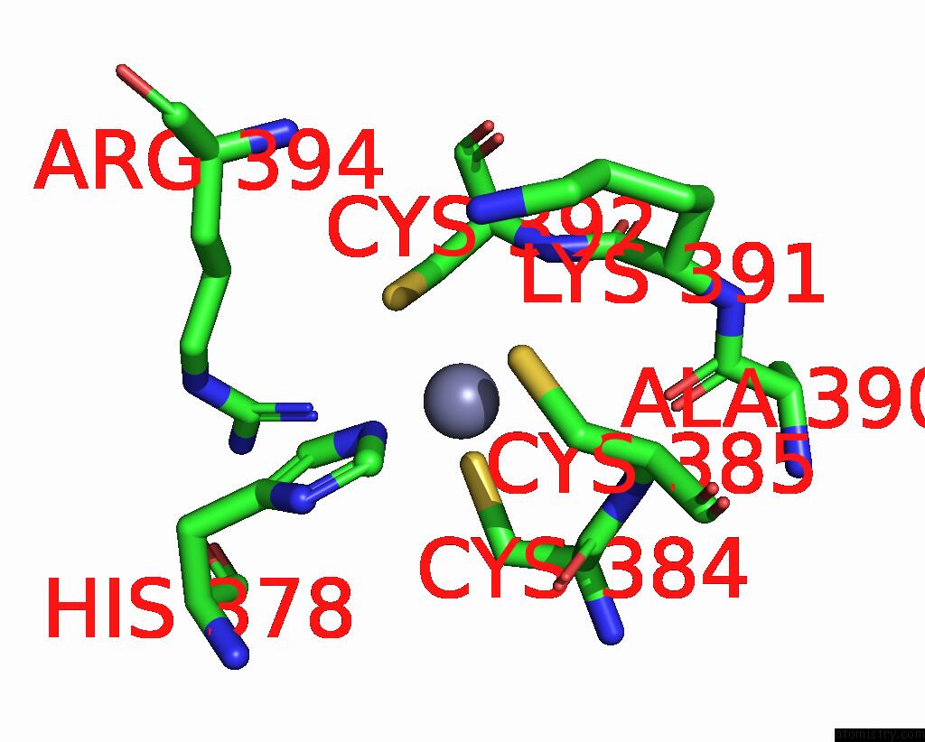
Zinc binding site 1 out of 6 in 5n6i
Go back to
Zinc binding site 1 out
of 6 in the Crystal Structure of Mouse Cgas in Complex with 39 Bp Dna

Mono view

Stereo pair view
Mono view
Stereo pair view
A full contact list of Zinc with other atoms in the Zn binding
site number 1 of Crystal Structure of Mouse Cgas in Complex with 39 Bp Dna within 5.0Å range:
|
Zinc binding site 2 out of 6 in 5n6i
Go back to
Zinc binding site 2 out
of 6 in the Crystal Structure of Mouse Cgas in Complex with 39 Bp Dna

Mono view

Stereo pair view
Mono view
Stereo pair view
A full contact list of Zinc with other atoms in the Zn binding
site number 2 of Crystal Structure of Mouse Cgas in Complex with 39 Bp Dna within 5.0Å range:
|
Zinc binding site 3 out of 6 in 5n6i
Go back to
Zinc binding site 3 out
of 6 in the Crystal Structure of Mouse Cgas in Complex with 39 Bp Dna

Mono view

Stereo pair view
Mono view
Stereo pair view
A full contact list of Zinc with other atoms in the Zn binding
site number 3 of Crystal Structure of Mouse Cgas in Complex with 39 Bp Dna within 5.0Å range:
|
Zinc binding site 4 out of 6 in 5n6i
Go back to
Zinc binding site 4 out
of 6 in the Crystal Structure of Mouse Cgas in Complex with 39 Bp Dna
Mono view

Stereo pair view
Mono view
Stereo pair view
A full contact list of Zinc with other atoms in the Zn binding
site number 4 of Crystal Structure of Mouse Cgas in Complex with 39 Bp Dna within 5.0Å range:
|
Zinc binding site 5 out of 6 in 5n6i
Go back to
Zinc binding site 5 out
of 6 in the Crystal Structure of Mouse Cgas in Complex with 39 Bp Dna

Mono view

Stereo pair view
Mono view
Stereo pair view
A full contact list of Zinc with other atoms in the Zn binding
site number 5 of Crystal Structure of Mouse Cgas in Complex with 39 Bp Dna within 5.0Å range:
|
Zinc binding site 6 out of 6 in 5n6i
Go back to
Zinc binding site 6 out
of 6 in the Crystal Structure of Mouse Cgas in Complex with 39 Bp Dna

Mono view

Stereo pair view
Mono view
Stereo pair view
A full contact list of Zinc with other atoms in the Zn binding
site number 6 of Crystal Structure of Mouse Cgas in Complex with 39 Bp Dna within 5.0Å range:
|
Reference:
L.Andreeva,
B.Hiller,
D.Kostrewa,
C.Lassig,
C.C.De Oliveira Mann,
D.Jan Drexler,
A.Maiser,
M.Gaidt,
H.Leonhardt,
V.Hornung,
K.P.Hopfner.
Cgas Senses Long and Hmgb/Tfam-Bound U-Turn Dna By Forming Protein-Dna Ladders. Nature V. 549 394 2017.
ISSN: ESSN 1476-4687
PubMed: 28902841
DOI: 10.1038/NATURE23890
Page generated: Sun Oct 27 22:37:27 2024
ISSN: ESSN 1476-4687
PubMed: 28902841
DOI: 10.1038/NATURE23890
Last articles
As in 1WMBAs in 1VHD
As in 1WCK
As in 1VHO
As in 1W2K
As in 1W0Y
As in 1TAD
As in 1TND
As in 1TYE
As in 1TZA