Zinc »
PDB 5mt3-5n31 »
5n1p »
Zinc in PDB 5n1p: Crystal Structure of the Polysaccharide Deacetylase BC1974 From Bacillus Cereus in Complex with N-Hydroxynaphthalene-1-Carboxamide
Protein crystallography data
The structure of Crystal Structure of the Polysaccharide Deacetylase BC1974 From Bacillus Cereus in Complex with N-Hydroxynaphthalene-1-Carboxamide, PDB code: 5n1p
was solved by
P.Giastas,
A.Andreou,
E.E.Eliopoulos,
with X-Ray Crystallography technique. A brief refinement statistics is given in the table below:
| Resolution Low / High (Å) | 48.45 / 1.45 |
| Space group | C 1 2 1 |
| Cell size a, b, c (Å), α, β, γ (°) | 196.103, 44.347, 99.132, 90.00, 98.83, 90.00 |
| R / Rfree (%) | 16.1 / 18 |
Other elements in 5n1p:
The structure of Crystal Structure of the Polysaccharide Deacetylase BC1974 From Bacillus Cereus in Complex with N-Hydroxynaphthalene-1-Carboxamide also contains other interesting chemical elements:
| Sodium | (Na) | 2 atoms |
Zinc Binding Sites:
The binding sites of Zinc atom in the Crystal Structure of the Polysaccharide Deacetylase BC1974 From Bacillus Cereus in Complex with N-Hydroxynaphthalene-1-Carboxamide
(pdb code 5n1p). This binding sites where shown within
5.0 Angstroms radius around Zinc atom.
In total 4 binding sites of Zinc where determined in the Crystal Structure of the Polysaccharide Deacetylase BC1974 From Bacillus Cereus in Complex with N-Hydroxynaphthalene-1-Carboxamide, PDB code: 5n1p:
Jump to Zinc binding site number: 1; 2; 3; 4;
In total 4 binding sites of Zinc where determined in the Crystal Structure of the Polysaccharide Deacetylase BC1974 From Bacillus Cereus in Complex with N-Hydroxynaphthalene-1-Carboxamide, PDB code: 5n1p:
Jump to Zinc binding site number: 1; 2; 3; 4;
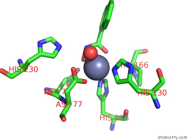
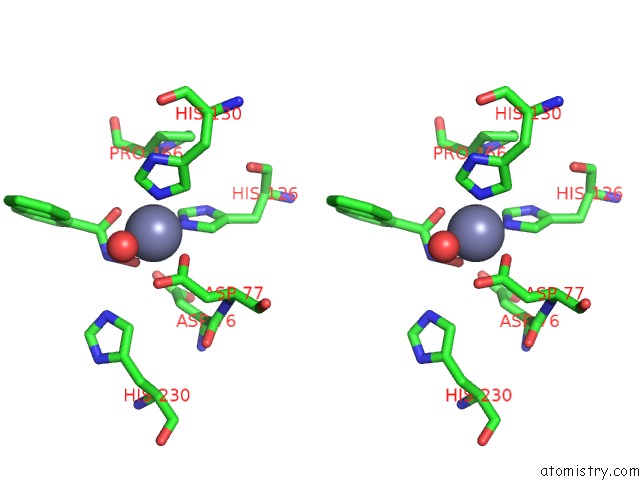
Zinc binding site 1 out of 4 in 5n1p
Go back to
Zinc binding site 1 out
of 4 in the Crystal Structure of the Polysaccharide Deacetylase BC1974 From Bacillus Cereus in Complex with N-Hydroxynaphthalene-1-Carboxamide
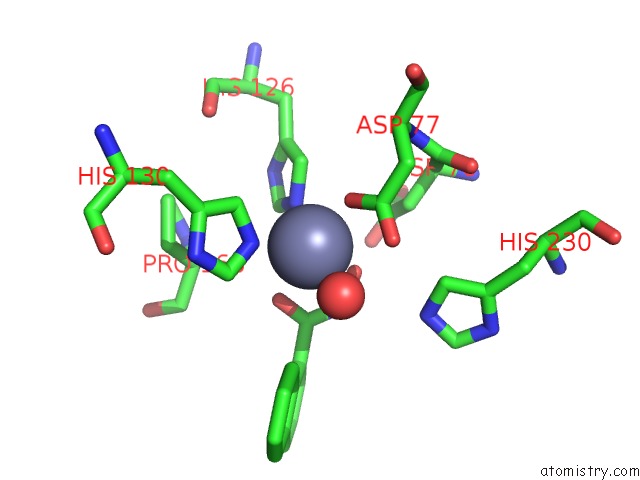
Mono view
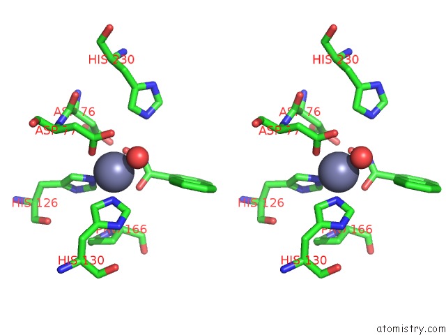
Stereo pair view
Mono view
Stereo pair view
A full contact list of Zinc with other atoms in the Zn binding
site number 1 of Crystal Structure of the Polysaccharide Deacetylase BC1974 From Bacillus Cereus in Complex with N-Hydroxynaphthalene-1-Carboxamide within 5.0Å range:
|
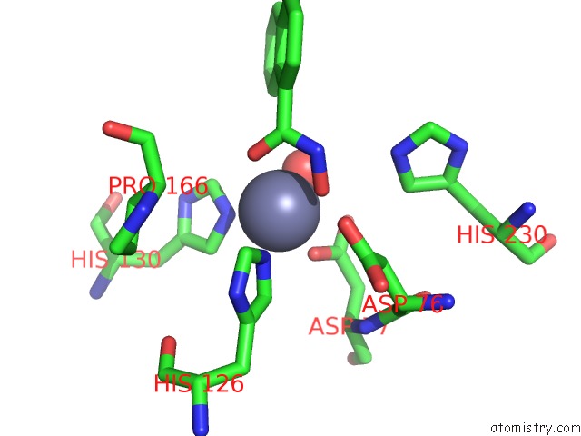
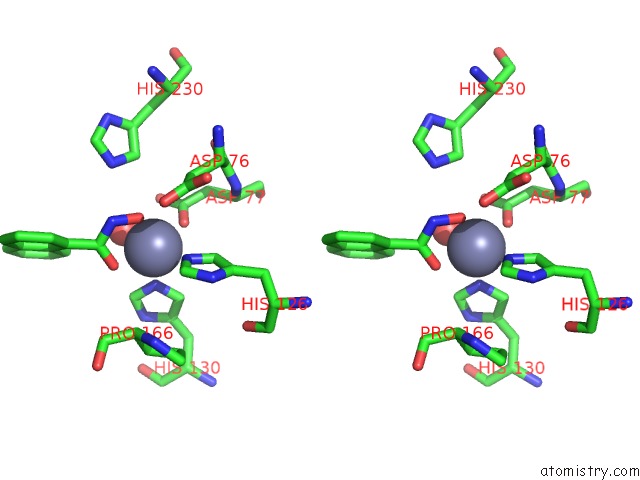
Zinc binding site 2 out of 4 in 5n1p
Go back to
Zinc binding site 2 out
of 4 in the Crystal Structure of the Polysaccharide Deacetylase BC1974 From Bacillus Cereus in Complex with N-Hydroxynaphthalene-1-Carboxamide
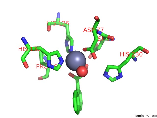
Mono view
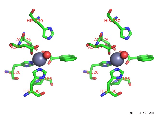
Stereo pair view
Mono view
Stereo pair view
A full contact list of Zinc with other atoms in the Zn binding
site number 2 of Crystal Structure of the Polysaccharide Deacetylase BC1974 From Bacillus Cereus in Complex with N-Hydroxynaphthalene-1-Carboxamide within 5.0Å range:
|
Zinc binding site 3 out of 4 in 5n1p
Go back to
Zinc binding site 3 out
of 4 in the Crystal Structure of the Polysaccharide Deacetylase BC1974 From Bacillus Cereus in Complex with N-Hydroxynaphthalene-1-Carboxamide
Mono view
Stereo pair view
Mono view
Stereo pair view
A full contact list of Zinc with other atoms in the Zn binding
site number 3 of Crystal Structure of the Polysaccharide Deacetylase BC1974 From Bacillus Cereus in Complex with N-Hydroxynaphthalene-1-Carboxamide within 5.0Å range:
|
Zinc binding site 4 out of 4 in 5n1p
Go back to
Zinc binding site 4 out
of 4 in the Crystal Structure of the Polysaccharide Deacetylase BC1974 From Bacillus Cereus in Complex with N-Hydroxynaphthalene-1-Carboxamide
Mono view
Stereo pair view
Mono view
Stereo pair view
A full contact list of Zinc with other atoms in the Zn binding
site number 4 of Crystal Structure of the Polysaccharide Deacetylase BC1974 From Bacillus Cereus in Complex with N-Hydroxynaphthalene-1-Carboxamide within 5.0Å range:
|
Reference:
P.Giastas,
A.Andreou,
A.Papakyriakou,
D.Koutsioulis,
S.Balomenou,
S.J.Tzartos,
V.Bouriotis,
E.E.Eliopoulos.
Structures of the Peptidoglycan N-Acetylglucosamine Deacetylase BC1974 and Its Complexes with Zinc Metalloenzyme Inhibitors. Biochemistry V. 57 753 2018.
ISSN: ISSN 1520-4995
PubMed: 29257674
DOI: 10.1021/ACS.BIOCHEM.7B00919
Page generated: Sun Oct 27 22:27:55 2024
ISSN: ISSN 1520-4995
PubMed: 29257674
DOI: 10.1021/ACS.BIOCHEM.7B00919
Last articles
Zn in 9MJ5Zn in 9HNW
Zn in 9G0L
Zn in 9FNE
Zn in 9DZN
Zn in 9E0I
Zn in 9D32
Zn in 9DAK
Zn in 8ZXC
Zn in 8ZUF