Zinc »
PDB 5mt3-5n31 »
5mwm »
Zinc in PDB 5mwm: Inositol 1,3,4,5,6-Pentakisphosphate 2-Kinase From M. Musculus in Complex with IP6
Enzymatic activity of Inositol 1,3,4,5,6-Pentakisphosphate 2-Kinase From M. Musculus in Complex with IP6
All present enzymatic activity of Inositol 1,3,4,5,6-Pentakisphosphate 2-Kinase From M. Musculus in Complex with IP6:
2.7.1.158;
2.7.1.158;
Protein crystallography data
The structure of Inositol 1,3,4,5,6-Pentakisphosphate 2-Kinase From M. Musculus in Complex with IP6, PDB code: 5mwm
was solved by
E.Franco-Echevarria,
J.Sanz-Aparicio,
B.Gonzalez,
with X-Ray Crystallography technique. A brief refinement statistics is given in the table below:
| Resolution Low / High (Å) | 57.46 / 2.60 |
| Space group | P 1 21 1 |
| Cell size a, b, c (Å), α, β, γ (°) | 60.550, 71.637, 61.822, 90.00, 111.66, 90.00 |
| R / Rfree (%) | 25.3 / 29 |
Zinc Binding Sites:
The binding sites of Zinc atom in the Inositol 1,3,4,5,6-Pentakisphosphate 2-Kinase From M. Musculus in Complex with IP6
(pdb code 5mwm). This binding sites where shown within
5.0 Angstroms radius around Zinc atom.
In total only one binding site of Zinc was determined in the Inositol 1,3,4,5,6-Pentakisphosphate 2-Kinase From M. Musculus in Complex with IP6, PDB code: 5mwm:
In total only one binding site of Zinc was determined in the Inositol 1,3,4,5,6-Pentakisphosphate 2-Kinase From M. Musculus in Complex with IP6, PDB code: 5mwm:
Zinc binding site 1 out of 1 in 5mwm
Go back to
Zinc binding site 1 out
of 1 in the Inositol 1,3,4,5,6-Pentakisphosphate 2-Kinase From M. Musculus in Complex with IP6
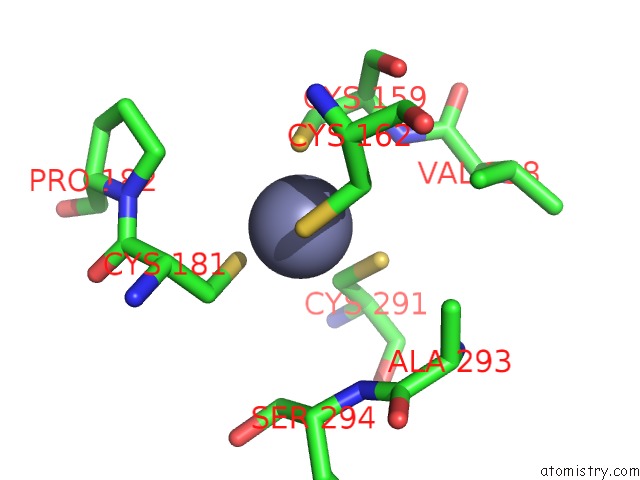
Mono view
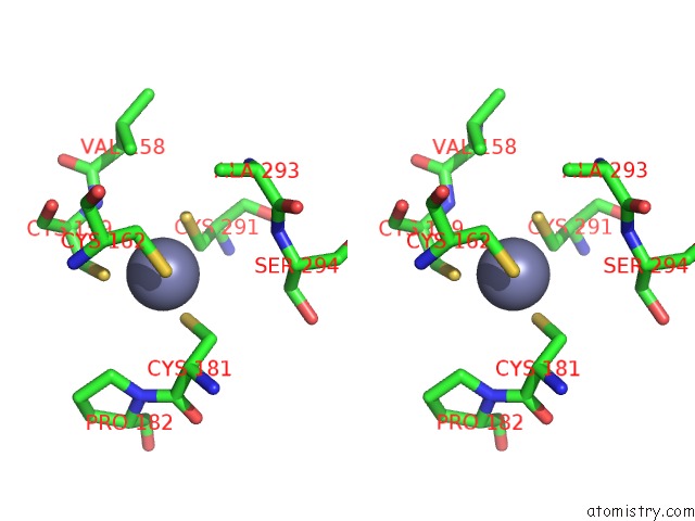
Stereo pair view
Mono view
Stereo pair view
A full contact list of Zinc with other atoms in the Zn binding
site number 1 of Inositol 1,3,4,5,6-Pentakisphosphate 2-Kinase From M. Musculus in Complex with IP6 within 5.0Å range:
|
Reference:
E.Franco-Echevarria,
J.Sanz-Aparicio,
C.A.Brearley,
J.M.Gonzalez-Rubio,
B.Gonzalez.
The Crystal Structure of Mammalian Inositol 1,3,4,5,6-Pentakisphosphate 2-Kinase Reveals A New Zinc-Binding Site and Key Features For Protein Function. J. Biol. Chem. V. 292 10534 2017.
ISSN: ESSN 1083-351X
PubMed: 28450399
DOI: 10.1074/JBC.M117.780395
Page generated: Sun Oct 27 22:22:49 2024
ISSN: ESSN 1083-351X
PubMed: 28450399
DOI: 10.1074/JBC.M117.780395
Last articles
Zn in 9MJ5Zn in 9HNW
Zn in 9G0L
Zn in 9FNE
Zn in 9DZN
Zn in 9E0I
Zn in 9D32
Zn in 9DAK
Zn in 8ZXC
Zn in 8ZUF